
Page 150 - IJB-8-2
P. 150
Laser printing of Gemcitabine on microneedles
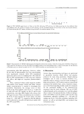
A B
Figure 6. HPLC-MS/MS quantification of Gem on the MNs following LIFT printing. (A) Table presenting the three different Gem
concentrations used, the number of repeats of LIFT printing, and the calculated amount of Gem found on the microneedle substrate.
(B) Graph showing the LIFT printed calculated dosage related to the nominal amount of Gem.
Figure 7. Representative LC-MS/MS chromatograms for Gem detected in mouse plasma, 15min after dosing mice with MNs (100 μg dose).
Upper panel: For the detection of Gem, the 264.1/112.0 MRM transition was used (retention time 1.6 min). Lower panel: for the detection
of Warfarin used as an internal standard, the 309.1/162.9 MRM transition was used (retention time 5.2 min).
concentrations after MN application. However, within 4. Discussion
60 min after IP administration, the Gem concentrations
were significantly reduced, while MN transdermal Current drug manufacturing techniques are performed
application led to a rise of circulating Gem. This indicates in specialized factories which follow best practice
that the MN administration of Gem may have reduced protocols for mass drug production. Digital printing
side effects and improved long-term pharmacokinetic techniques can be used in medical facilities (i.e.,
profile. hospitals, medical centers) to maximize the advantage of
Thus, a sustained release profile might be possible drug personalization. LIFT method requires a specialized
with smaller Gem dosages loaded onto MNs. Indeed, equipment that includes laser, optics, translation stages,
further experimentation is needed to investigate this and a robot-based pipetting module (under development)
possibility. Importantly, the average levels of Gem for the automated loading of the pharmaceutical
levels in the mouse plasma were found at 1.8 μM and compound onto the donor substrate. The cost of LIFT
4.1 μM (479 and 1087 ng/mL) for the 15 and 60 min method depends on the automation level and throughput
respectively, even when MNs loaded with a lower dose when it comes to the mass production. It offers advantages
(100 μg) were used. These concentrations are well above such as the ability to use any potency and viscosity drug
the IC50 of Gem reported for several cancer cell lines formulations, adaptable resolution (10 – 500 μm) for
(range 20 – 1000 nM [30-33] ), suggesting that the efficacy coating any kind of thin-film substrates including MN
can be achieved in clinically relevant cancer models with patches- as well as individual MNs- and minimal drug
the LIFT-coated MNs. formulation waste.
142 International Journal of Bioprinting (2022)–Volume 8, Issue 3