
Page 15 - IJB-8-3
P. 15
Liu, et al.
Table 1. Quantified physical properties of the PET membrane and EHDJ-printed ultrathin scaffolds (n=3)
Thickness Pore size Pore density Porosity Fiber TEER
(μm) (μm) (pores/cm ) (%) diameter (μm) (Ω·cm )
2
2
PET membrane 13.63±1.37 0.34±0.15 4.0×10 6[25] 1.85±0.55 - 73.70±0.71
S20 7.88±0.92 22.91±2.16 6.25×10⁴ 33.00±4.74 20.50±0.88 63.14±1.44
S50 7.96 ± 0.74 52.17 ± 1.27 2.04 × 10⁴ 55.04 ± 3.46 21.23 ± 1.27 50.26 ± 1.17
A It is also important for scaffolds to have similar
biomechanical properties to Bruch’s membrane to avoid
incompatibility with surrounding tissues in vivo. In the
human body, Young’s modulus of Bruch’s membrane
ranges from 1.0 to 18.8 MPa . In contrast, Young’s
[26]
modulus of the commercial PET membrane is about
10 times higher (~180 MPa) , making it too stiff
[27]
and susceptible to damage in the surrounding tissue
if transplanted. The Young’s modulus for S20 and S50
B scaffolds are much lower, that is, 45.5 ± 5.3 MPa and
8.9 ± 3.2 MPa, respectively (Table 2). In addition, the
ultimate tensile stress, ultimate tensile strain, yield stress,
and yield strain of the S20 and S50 were significantly
different (Figure 4A and B). Since large pore scaffolds
require lesser fibers, S50 scaffold is much easier to
deform, whereas S20 can withstand higher stress. Both
scaffolds have similar elastic properties compared with
C the actual Bruch’s membrane. Young’s moduli of S50 are
within the range of Bruch’s membrane and thus might be
better suited for implant applications.
Moreover, both PCL scaffolds have a good
degradation behavior under lipase treatment (Figure 4C),
with ~40% of the scaffolds being degraded after 48 h
of immersing with lipase solution in 37°C (calculated
by Equation 2.2). In contrast, the commercial PET
membrane is not biodegradable. Thus, the PCL scaffolds
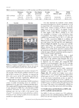
Figure 3. Comparison of morphological characteristics of EHDJ- may be promising for culturing RPE monolayers, with
printed PCL scaffolds and PET membrane. The top view and side potential application in transplantation.
view of (A) PET membrane, (B) S20, and (C) S50 under SEM. Both PET (materials used to make commercial
membrane) and PCL are hydrophobic. PET membrane
membrane has an extremely low porosity at around 1.5%. exhibited a water contact angle of 62.28 ± 1.07°
The porosity of the S50 PCL scaffold is around 55% and (Figure 4D), S20 scaffold has a higher water contact
that of S20 is around 33%. Therefore, we expected the angle of 95.99 ± 1.72°, and the S50 has the highest water
RPE cells to grow on the PET membrane and that the contact angle of 112.81 ± 1.66°. The connective tissues
PCL scaffold would have significantly different mass in vivo have higher hydrophilicity as they are made of
exchange behavior with the culture media. proteins, but the hydrophobicity for PCL scaffolds is an
The thickness of the membranes for RPE monolayer essential factor as a substrate for much small monolayer
culture is another determining factor for suitability of cell RPE to grow on without falling through the pores (20
implanting in vivo since there is limited subretinal space. and 50 μm). The water on the hydrophobic scaffold can
The commercial PET membrane has a thickness of around form a layer of water film and prevent cells from falling.
13 μm, measured by SEM. Due to the limitation of PET
membrane fabrication, the actual values of membrane 3.3. Proliferation and distribution of RPE cells
thickness may vary up to 60% of the nominal value on PCL scaffolds and membrane
(10 μm) . In contrast, the printed scaffold has a uniform The ARPE-19 cells were cultured over 70% confluency
[25]
thickness of 7 μm, making it suitable for in vivo study and and inoculated to the scaffolds/membrane. ARPE-19 cells
possible for implant purpose . (94%) have cell sizes ranging from 9 to 20 μm with mean
[2]
7 International Journal of Bioprinting (2022)–Volume 8, Issue 3