
Page 176 - IJB-8-3
P. 176
Composite Scaffolds for Skin Repair
range of 3 – 8 kPa [31-34] . It should be emphasized that the A B
photoinitiator concentration, degree of methacrylation,
UV intensity, and exposure time can affect the mechanical
properties of GelMA. Zhang et al. summarized
the effects of different additives on the mechanical
properties of GelMA hydrogel in their review article.
They claimed that most of the additives could improve
the mechanical properties of the GelMA hydrogel [35] . It
has been reported that the DE microparticles could serve
as reinforcing filler in hydrogel networks [20,21] . Based
on above, the mechanical properties of DE-Gel will be
improved by the incorporated DE microparticles.
3.3. Biological activities of the 3D-printed
DE-containing scaffolds in vitro
The biological effects of DE incorporated scaffolds
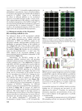
on HDFs and HUVECs were evaluated separately. Figure 3. Evaluation of viability of HDFs in the scaffolds. The
CLSM images demonstrated the stained nuclei and distribution of HDFs seeded on 3D-printed scaffolds with various
cytoskeleton of HDFs and HUVECs seeded on the concentrations of DE microparticles for (A) 1 and (B) 5 days using
3D-printed scaffolds. Although HDFs presented uneven CLSM observation. Scale bar: 500 μm.
distribution as aggregated clumps on the scaffolds on
day 1 (Figure 3A), after 5 days of culture, HDFs rapidly
proliferated, migrated, and covered the entire surface of A B
the scaffolds (Figure 3B). Besides, the cell proliferation
assay was also performed after 1, 3, and 5 days. The
HDFs adhered on the 3D-printed scaffolds in each
group performed great proliferation independent of DE
concentrations (Figure 4A).
Interestingly, the HUVECs seeded on DE- C D
containing scaffolds exhibited better cell spreading
with more obvious presence of filopodia than those on
GelMA scaffolds on the 1 day (Figure 5A). Compared
st
with HDFs, HUVECs appeared more sensitive that the
cell viability was significantly affected by the changes
of DE content. According to Figure 5C and D, the
cell numbers of HUVECs adhering on the 5DE-Gel
and 10DE-Gel scaffolds were higher than that in Gel Figure 4. Proliferation and differentiation of cells in the 3D-printed
group, and the incorporation of DE greatly increased DE-Gel composite scaffolds. Proliferation behaviors of (A) HDFs
the cell spreading area of HUVECs. Among the five and (B) HUVECs on the Gel, 5DE-Gel, 10DE-Gel, and 20DE-Gel
groups, the Gel, 5DE-Gel, 10DE-Gel, and 20DE- scaffolds for 1, 3, and 5 days (n = 4). (C) The expression levels of
Gel scaffolds were able to support the cell survival genes related to angiogenesis in HUVECs on 3D-printed scaffolds
and spreading during culture for 5 days (Figure 5B). containing different concentrations of DE microparticles (n = 3).
It is worth noting that the Gel, 5DE-Gel, and 10DE- (D) Cumulative Si ion released from the scaffolds with different
concentrations of DE after culturing in cell culture condition for
Gel scaffolds presented good performance in network 5 days (n = 3). *P < 0.05, **P < 0.01, and ***P < 0.001.
formation of HUVECs. Especially, the 5DE-Gel
scaffolds significantly promoted HUVECs proliferation increased the roughness of scaffolds, thereby providing
as shown in Figure 4B. In contrast, the 30DE-Gel binding sites for orientation and movement of cells
group negatively affected cell viability, resulted in poor [38]
cell state. The reason might be that high contents of adhering on the surface . Therefore, the satisfactory
DE could release excess amount of ion which triggered attachment, migration, and proliferation of skin cells
adverse side effects on cell activities. As known, the cell laid a solid foundation for the application of scaffolds
attachment and spreading always strongly influenced in wound therapy.
by the surface topography [36,37] . Fortunately, the Subsequently, the angiogenesis activity of 3D-printed
incorporation of proper amount of DE microparticles DE-containing scaffolds was detected by RT-qPCR. It is
168 International Journal of Bioprinting (2022)–Volume 8, Issue 3