
Page 179 - IJB-8-3
P. 179
Ma, et al.
A B burn wound healing in vivo. Due to the stimulation of
Si ion to HUVECs, rapid sprouting and rebuilding of
vasculature were activated [45,62,63] , and then, the skin
cells could be supplied with sufficient nutrients and
oxygen [62,64] , contributing to the high speed of wound
closure in 5DE-Gel group. Therefore, the 3D-printed
DE-containing composite scaffolds exerted prominent
repair effects on burn wound healing and skin tissue
regeneration.
C D 4. Conclusions
In this study, we developed a 3D-printed composite
scaffold with the incorporation of DE microparticles,
which could serve as a functional dressing for the
treatment of skin burn wound. Natural DE microparticles
with porous morphology could be used as a stable Si
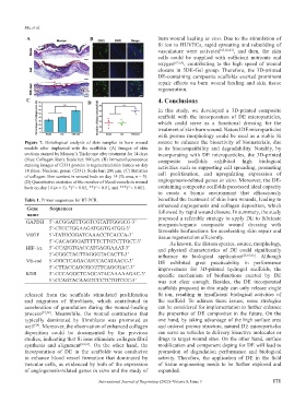
Figure 7. Histological analysis of skin samples in burn wound source to enhance the bioactivity of biomaterials, due
models after implanted with the scaffolds. (A) Images of skin to its biocompatibility and degradability. Notably, by
sections stained by Masson’s Trichrome after treatment for 14 days incorporating with DE microparticles, the 3D-printed
(blue: Collagen fiber). Scale bar: 500 μm. (B) Immunofluorescence composite scaffolds exhibited high biological
staining images of CD31 protein in regenerated skin tissues on day activities such as supporting cell spreading, promoting
14 (blue: Nucleus, green: CD31). Scale bar: 200 μm. (C) Statistics
of collagen fiber content in wound beds on day 14 (% area, n = 5). cell proliferation, and upregulating expression of
(D) Quantitative statistics of the number of blood vessels in wound angiogenesis-related genes in vitro. Moreover, the DE-
beds on day 14 (n = 5). *P < 0.05, **P < 0.01, and ***P < 0.001. containing composite scaffolds possessed ideal capacity
to create a bionic environment that efficaciously
Table 1. Primer sequences for RT-PCR. benefited the treatment of skin burn wounds, leading to
enhanced angiogenesis and collagen deposition, which
Gene Sequences
name followed by rapid wound closure. In summary, the study
GAPDH 5’-ACGGATTTGGTCGTATTGGGCG-3’ proposed a referable strategy to apply DE to fabricate
inorganic/organic composite wound dressing with
5’-CTCCTGGAAGATGGTGATGG-3’ favorable biofunctions for accelerating skin repair and
VEGF 5’-TATGCGGATCAAACCTCACCA-3’ tissue regeneration efficiently.
5’-CACAGGGATTTTTCTTGTCTTGCT-3’ As known, the diatom species, source, morphology,
HIF-1α 5’-CCATGTGACCATGAGGAAAT-3’ and physical characteristics of DE could significantly
5’-CGGCTAGTTAGGGTACACTT-3’ influence its biological application [23,65,66] . Although
VE-cad 5’-GGCTCAGACATCCACATAACC-3’ DE exhibited great practicability in performance
5’-CTTACCAGGGCGTTCAGGGAC-3’ improvement for 3D-printed hydrogel scaffolds, the
KDR 5’-CCCAGGCTCAGCATACAAAAAGAC-3’ specific mechanism of biofunctions exerted by DE
5’-CCAGTACAAGTCCCTCTGTCCC-3’ was not clear enough. Besides, the DE incorporated
scaffolds proposed in this study can only release single
released from the scaffolds stimulated proliferation Si ion, resulting in insufficient biological activities of
and migration of fibroblasts, which contributed to the scaffold. To address these issues, some strategies
acceleration of granulation during the wound-healing can be considered for implementation to further enhance
process [57,58] . Meanwhile, the wound contraction that the properties of DE composites in the future. On the
typically dominated by fibroblasts was promoted as one hand, by taking advantage of the high surface area
well [59] . Moreover, the observation of enhanced collagen and ordered porous structure, natural DE microparticles
deposition could be documented by the previous can serve as vehicles to delivery bioactive molecules or
studies, indicating that Si ions stimulate collagen fibril drugs to target wound sites. On the other hand, surface
synthesis and alignment [60,61] . On the other hand, the modification and component doping for DE will lead to
incorporation of DE in the scaffolds was conducive promotion of degradation performance and biological
to enhance blood vessel formation that dominated by activity. Therefore, the application of DE in the field
vascular cells, as evidenced by both of the expression of tissue engineering needs to be further explored and
of angiogenesis-related genes in vitro and the study of expanded.
International Journal of Bioprinting (2022)–Volume 8, Issue 3 171