
Page 110 - IJB-8-4
P. 110
Effect of Bioprinting-Associated Shear Stress and Hydrostatic Pressure
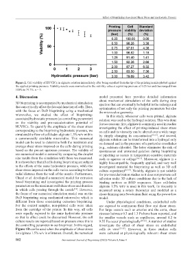
Figure 2. Cell viability of HUVECs in alginate solution immediately after being expelled from the tip of the printing nozzle plotted against
the applied printing pressure. Viability results were normalized to the viability value at a printing pressure of 0.25 bar and thus ranged from
100% to 79.7%. n = 3.
4. Discussion model presented here provides detailed information
about mechanical stimulation of the cells during drop
3D bioprinting is accompanied by mechanical stimulation ejection that can eventually be helpful in the redesign and
that can critically affect the fate and function of cells. Here, optimization of not only the printing parameters but also
with the focus on DoD bioprinting using a mechanical the microvalve geometry.
microvalve, we studied the effect of bioprinting- In this study, whenever cells were printed, alginate
associated hydrostatic pressure (as controlling parameter) solution was used as the hydrogel solution. This was done
on the viability and pre-vascularization potential of for two reasons: first, alginate is commonly used in studies
HUVECs. To quantify the amplitude of the shear stress investigating the effect of printing-induced shear stress
corresponding to the bioprinting hydrostatic pressure, we on cells and its viscosity can be altered over a wide range
simulated the flow of cell-laden alginate 1.5% w/v within by simply changing its concentration [10,13] , and second,
a commercially available microvalve. This numerical alginate solution can be transformed into a hydrogel only
model can be used to determine both the maximum and on demand and in the presence of a particular crosslinker
average shear stress imposed on the cells during printing (e.g., calcium chloride). The latter eliminates the risk of
based on the pre-set upstream pressure. The validity of spontaneous and unwanted gelation during bioprinting
our numerical model is assured by comparing the droplet or pipetting as occurs in temperature-sensitive materials
size results from the simulation with those we measured. such as agarose or collage [14,15] . Moreover, alginate is a
It is shown here that all cells during bioprinting are subject highly biocompatible, frequently applied, and very well
to the effects of the same hydrostatic pressure, while the investigated material for bioprinting as well as 3D cell
shear stress imposed on the cells varies according to their culture experiment [16-19] . Notably, alginate is not suitable
radial distance from the wall of the nozzle. Furthermore, for pre-vascularization studies as it does not promote cell
Chand et al. developed a numerical model for extrusion adhesion under 3D culture conditions due to the lack of
based bioprinting and investigated the printing process binding motives as RGD sequences. Since cell-laden
parameters on the maximum wall shear stress and duration alginate 1.5% wt/v is used in this work, its viscosity is
in which cells passing through the nozzle . However, measured using a rotary rheometer and modeled as a
[12]
the focus of our numerical simulation model was on the shear-thinning non-Newtonian fluid using the power-law
DoD bioprinting using mechanical microvalve which is model.
different from those considering extrusion bioprinting. Under physiological conditions, endothelial cells
For the control samples, non-printed cells were taken are exposed to continuous fluid flow and shear stress.
from the cartridge of the printer. In this way, all cells For large vessels such as arteries and the aorta, shear
were equally exposed to the same hydrostatic pressure stresses between 0.3 and 1.3 Pa have been reported, and
so that its effect could be discounted. However, the cell for smaller vessels such as capillaries, around 4.2 to
culture results are reported based on pressure since this is 9.55 Pa occur physiologically [20-22] . A number of studies
the controlling parameter in our bioprinter. In this regard, investigate the effect of shear stress on endothelial
Figure 1B can be used when the amplitude of shear stress cells in vitro [23-25] . However, in those studies cells
for alginate 1.5% w/v is of interest. Overall, the numerical were cultured at physiologically relevant shear stress
102 International Journal of Bioprinting (2022)–Volume 8, Issue 4