
Page 126 - IJB-9-1
P. 126
International Journal of Bioprinting Fabrication of 3D breast tumor model for drug screening
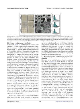
Figure 3. Structure of 3D-printed Gel/SA/dECM scaffolds. 5G3S1d and 6G3S1d were printed with the temperature of 20° and pressure of 0.20 Mpa, while
7G2S1d was printed with the temperature of 24° and pressure of 0.24 Mpa. (A) Macrostructure of the scaffolds before and after crosslinking by 3% CaCl 2
and EDC/NHS. Scale bar: 5 mm. (B) Microstructure of crosslinked scaffolds observed by scanning electron microscope. The left images (scale bar: 100 μm)
were enlarged and shown on the right (scale bar: 100 μm). (C) Distribution of small pore size in the scaffolds.
3.3. Structure and pore size of scaffolds size in the scaffold turned out to be 20–60 μm, while the
As shown in Figure 3A, the three printed scaffolds were large pore size was 600–1000 μm, and the small pore size
translucent with high resolution and clear pore structure, increased as large pore size increased. In contrast, the
but the structure was loose and easy to deform with three groups of 3D-printed scaffolds in our study had
external forces. In order to further improve the stability smaller large pores but larger small pores, indicating that
of the scaffolds, CaCl and chemical crosslinking agent the addition of gelatin and sodium alginate improved the
2
EDC/NHS were both used to crosslink the scaffolds. After porosity of the material and was conducive to providing
crosslinking, the stiffness and resolution of the scaffolds support for the growth of more cells.
were further improved. After freeze-drying, the scaffolds 3.4. Physical properties and hemolytic properties of
presented a white slice with evenly distributed pore scaffolds
structure. In order to further observe the microstructure IR spectra of the scaffold before and after crosslinking
of the scaffolds, they were observed by SEM. Figure 3B were shown in Figure 4A and B. It could be seen that
showed a series of uniformly distributed macropores which the main absorption peaks before and after crosslinking
were formed from printing gaps and many interconnected showed no significant difference, but the transmittances
micropores on the beam which were created by of some absorption peaks were changed. This was due to
lyophilization after crystallization. However, there were no CaCl is a physical crosslinking agent, combining alginate
pore structures in some areas, while the surface exhibited and divalent cation to form a stable network structure by
2
rough ravine, similar to the results of Chaji et al. . The ionic bond, thus improving the stability of the system.
[55]
complex network of small pores nested with large holes This process does not change the chemical composition
within the scaffold increased the specific surface area of of the material . In addition, EDC/NHS is a chemical
[58]
the scaffold and reduced the risk of dissolution in liquid crosslinking agent, whose crosslinking mechanism is to
environment . The large pore sizes of the three groups of transform the amino and carboxyl groups in the system
[56]
scaffolds were 262.62 ± 49.78 μm, 202.57 ± 14.23 μm and into amide bonds, which can repair collagen broken
533.58 ± 52.41 μm, respectively. The pore size distribution in the process of decellularization . Therefore, in the
[49]
was shown in Figure 3C. The small pore sizes of the three infrared spectrogram after crosslinking, the transmittance
groups of scaffolds ranged from 15.97 μm to 140.32 μm, of absorption peaks C=O and N-H changed greatly, and
22.55 μm to 173.67 μm, and 5.10 μm to 50.21 μm, the two absorption peaks at 1653 cm and 1553 cm were
–1
–1
respectively. The complex porous structure with these pore the same as those of gelatin, corresponding to the C=O
sizes could promote the growth, adhesion, and invasion of stretching vibration of amide I and N-H deformation
cells on the scaffold, and the large pore size was beneficial vibration of amide II and C=N in protein, respectively [59,60] .
to improve the exchange efficiency of metabolic waste and In addition, N-H stretching vibration peaks of amide I
nutrients in the tumor model.
and amide II were observed at 3294 cm and 3086 cm ,
−1
−1
Jeong et al. prepared porous scaffolds by 3D printing respectively. N-deformation vibration peak of amide III
[57]
using pure porcine liver dECM as bioink. The small pore was observed at 1238 cm , and absorption peak of GAG
−1
Volume 9 Issue 1 (2023) 118 https://doi.org/10.18063/ijb.v9i1.630