
Page 130 - IJB-9-1
P. 130
International Journal of Bioprinting Fabrication of 3D breast tumor model for drug screening
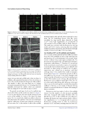
Figure 6. Infiltration of L929 within the Gel/SA/dECM scaffolds was observed by laser scanning confocal microscope. (A) 3D views of cells grown in the
scaffolds at 1 d, 4 d, and 7 d (scale bar: 500 μm). (B) Distribution of cells on different depth in 6G3S1d scaffolds (scale bar: 500 μm).
increased significantly, and cell clusters appeared in some
areas. The cell morphology became more flat, which
resembled the long spindle shape observed by Kwak
et al. . Under high magnification, the pseudopodia of
[71]
cells attached to the scaffolds could be clearly observed.
This result was consistent with the fluorescence staining
results and CCK-8 results, indicating that our scaffolds
exhibited good biocompatibility, and cell clusters with high
viability could be formed after 7 d of inoculation.
3.6. Viability of 4T1 on 3D scaffolds and 2D plates
4T1 is a tumor cell line extracted by Fred Miller and others
from mice with portability as well as high tumorigenicity
and invasion, and could metastasize from primary breast
tumors to distant tissues and organs spontaneously. The
invasion and metastasis of 4T1 are identical to metastatic
breast tumor cells of human . Therefore, 4T1 was utilized
[72]
in the present study as seed cells to construct the metastatic
tumor model. Mice metastatic breast cancer cells 4T1 were
inoculated on three groups of scaffolds and 2D plates,
Figure 7. Obvious growth of L929 cells on Gel/SA/dECM scaffolds at 1 respectively, and cultured in an incubator with fluid
d and 7 d as observed by a scanning electron microscope. Cell colonies exchange every 2 d to establish the model of metastatic
were observed at 7 d. The images in upper panel (scale bar: 100 μm) were
enlarged and shown in the lower panel (scale bar: 10 μm) to show the breast cancer. Figure 8A–C showed the growth of cells on
antennae binding cells with the scaffolds. the scaffold on 1 d, 4 d, and 7 d of culture. Hoechst could
travel across the cell membrane and bind to small grooves
wastes between internal scaffold, and culture medium. In on the DNA double strands in living cells, and emit blue
addition, cells penetrated the interior from the surface, so fluorescence at an excitation wavelength of 461 nm. So,
fewer cells could be found at the deeper site of the scaffold. it is often used to label living cells. However, at the same
The number of cells at the same depth on 4 d culture time, it can also bind with gelatins and other biocompatible
increased compared with that on 1 d, indicating that L929 scaffolds to fluoresce, so the results of Hoechst staining
gradually penetrated the scaffold, which was consistent should be compared with those of Calcein-AM staining to
with the change of 3D view with the days of culture. draw a conclusion.
The growth morphology of cells on the scaffolds was Fluorescence staining results of cells on the scaffolds
observed by SEM, and the results were shown in Figure 7. showed that the number of cells increased gradually
It can be seen that the cells were mostly attached to the with time. Compared with mouse fibroblast L929, the
corner of the scaffold, the ravine or the edge of the large distribution of 4T1 on the scaffold was more dispersed,
pores. On 1 d, several cells, which were almost spherical, forming cell clusters and tumor spheres clearly visible at
were observed on the scaffolds. A few cells were distributed low magnification on 7 d. Compared with the previous
together while most of them were randomly scattered in fluorescence staining results of L929, the fluorescence
the same area. On 7 d, the number of cells on the scaffolds intensity of 4T1 was lower, resulting from the fewer 4T1
Volume 9 Issue 1 (2023) 122 https://doi.org/10.18063/ijb.v9i1.630