
Page 133 - IJB-9-1
P. 133
International Journal of Bioprinting Fabrication of 3D breast tumor model for drug screening
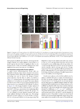
Figure 11. Progression of 4T1 tumor spheres on the Gel/SA/dECM scaffolds. (A) Confocal laser microscopy was used to observe the progression of tumor
spheres on the three groups of scaffolds for 1 d, 4 d, and 7 d (scale bar: 200 μm). (B) Growth and morphology of 4T1 in 2D environment (scale bar: 200
μm). (a), (b), (c), and (d) show the images of 4T1 under optical microscope on 1 d, 3 d, 5 d, and 7 d, respectively. (C) Quantitative detection of the change
of tumor sphere diameter on the scaffolds on 3 d, 5 d, and 7 d. The diameters were evaluated by IPP software. *P<0.05, **P<0.01 represent significant
difference between two groups.
three groups of scaffolds was observed, indicating that the diameters of some tumor spheres were still small, similar
change of gelatin and sodium alginate concentration in a to that on 3 d. It was speculated that these spheres may
small range in this study had no significant effect on the be formed by newly proliferating cells during culture. On
invasion of 4T1. If the concentration difference was more 7 d, the average diameters of tumor spheres increased to
significant, the results may turn out to be different. 116.19 ± 4.63 μm, 148.52 ± 8.02 μm, and 122.25 ± 7.63 μm,
respectively. There were significant differences between the
3.8. Formation and growth of tumor spheres groups, indicating that the tumor spheres became mature
In native tumor microenvironment, tumor cells can at this time, showing the influence of scaffold materials on
gradually develop into tumor spheres. In order to determine tumor cell viability and tumorigenicity.
the progression of 4T1 cells on the scaffolds, laser confocal
microscopy was used to observe the change of tumor Overall, tumor spheres on the 6G3S1d scaffolds were the
sphere diameter over the time of culture. As shown in largest in diameter and grew at a faster rate than those on
Figure 11A, scattered tumor spheres began to form on the the other two groups, consistent with the results of viability.
scaffolds on 3 d, but the size was small. The tumor spheres However, from 3 d to 7 d, the growth rate of tumor spheres
in different areas of the same group were photographed and on the same group of scaffolds remained almost the same,
their diameters were measured by IPP software. As shown indicating that the proliferation rate of tumor spheres on the
in Figure 11C, the average diameters of tumor spheres of scaffolds was relatively stable. The shape and size of spheres
the three groups were 78.81 ± 6.06 μm, 88.91 ± 12.61 μm, formed by 4T1 cells on the scaffolds were similar to the results
[74]
and 84.87 ± 10.93 μm, respectively, which indicating there obtained by Keklikoglou et al. . Lv et al. established a
[56]
were no significant difference between the groups. On 3D-printed chitosan/gelatin scaffold, which was inoculated
5 d, the diameter of tumor spheres increased and more with human breast cancer cells MCF-7, and the diameter of
tumor spheres developed in the same area. The average spheres was about 150 μm after 7 d, which was close to that of
diameters of tumor spheres on 5 d were 96.99 ± 6.55 μm, 6G3S1d in the present study. Bright field images showed that
115.18 ± 4.29 μm, and 100.06 ± 14.43 μm, respectively. The tumor spheres were attached at the corner of the scaffolds and
Volume 9 Issue 1 (2023) 125 https://doi.org/10.18063/ijb.v9i1.630