
Page 132 - IJB-9-1
P. 132
International Journal of Bioprinting Fabrication of 3D breast tumor model for drug screening
Figure 9. Morphology of 4T1 cells cultured on the Gel/SA/dECM scaffolds for several days. Pseudopodia were observed clearly on 1 d, and cell colonies
appeared on the scaffolds after 4 d of culture (4 d and 7 d). The images on the right panel (scale bar: 10 μm) show magnified images of those on the left
panel for each day category (scale bar: 100 μm).
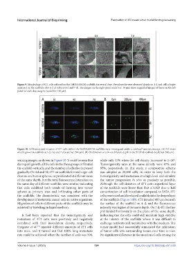
Figure 10. Infiltration and invasion of 4T1 cells within the Gel/SA/dECM scaffolds were investigated under a confocal laser microscope. (A) 3D views
of cells grown in scaffolds at 1 d, 4 d, and 7 d (scale bar: 500 μm). (B) Distribution of cells on different depth in the 5G3S1d scaffolds (scale bar: 500 μm).
6
staining images, as shown in Figure 10. It could be seen that while only 45% when the cell density increased to 1×10 .
during cell growth, all the cells in the three groups infiltrated Tumorigenicity rates at the same density were 87% and
the scaffold vertically, and the number of cells also increased 95%, respectively. In this study, a compromise scheme
gradually. On 4 d and 7d, 4T1 on scaffolds formed larger cell was adopted as 20,000 cells, in order to keep both the
clusters and tumor spheres, or proliferated at different areas tumorigenicity and metastasis at a high level and simulate
of the same depth, but the total fluorescence intensities on the tumor progression in vivo as precisely as possible.
the same day of different scaffolds were similar, indicating Although the cell densities of 4T1 onto superficial layer
that cells exhibited both trends of forming into tumor of the scaffolds were lower than that of L929 due to half
spheres at primary sites and infiltrating other parts of concentration of cell inoculation compared to L929, 4T1
the scaffolds. The characteristic was consistent with the cells penetrated and developed availably into the deeper layer
development of metastatic cancer cells in native organisms. of the scaffolds (Figure 10B). 4T1 invaded 400 μm beneath
Migration of cells to different parts of the scaffolds may be the surface of the scaffold on 4 d, and the fluorescence
achieved by travelling in liquid medium. intensity was higher at the same depth. On 7 d, 4T1 further
proliferated horizontally on the plane of the same depth,
It had been reported that the tumorigenicity and indicating that the cells could still maintain high viability
metastasis of 4T1 cells were positively and negatively at the interior of the scaffolds where it was difficult to
correlated with their inoculation density, respectively. exchange nutrients and metabolites with the medium. The
[73]
Gregorio et al. injected different amounts of 4T1 cells tumor model had successfully stimulated the infiltration
into mice, and it turned out that 100% lung metastasis of tumor cells into surrounding tissues over time in vivo.
rate could be achieved when the number of cells was 500, No significant difference in the invasion of cells among the
Volume 9 Issue 1 (2023) 124 https://doi.org/10.18063/ijb.v9i1.630