
Page 127 - IJB-9-1
P. 127
International Journal of Bioprinting Fabrication of 3D breast tumor model for drug screening
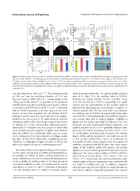
Figure 4. Physical characteristics of the 3D-printed Gel/SA/dECM scaffolds. (A) FITR spectra of the scaffolds before and after crosslinking. (B) FITR
spectra at 1000–1800cm . (C) Swelling ratio of the scaffolds. (D) Swelling ratio during the initial 6 h. (E-G) Water contact angles of the scaffolds at 0 s.
–1
(H) Water contact angles of the scaffolds at 0 s, 0.5 s, and 1 s. **P<0.01 represents significant difference between two groups. (I) Stress-strain curve of the
scaffolds. (J) Compressive modulus of the scaffolds within elastic deformation stage. **P<0.01 represents significant difference between two groups. (K)
Porosity of the scaffolds was investigated. NS represents no significant difference between two groups. Hemolytic test was performed on scaffolds and result
is shown in (L).
was also observed at 1048 cm −1[59,61] . The absorption peak while increased slowly after 2 h, and gradually stabilized
at 1390 cm was the stretching vibration of C-N, and after 48 h. After 72 h, the swelling ratios of 5G3S1d,
–1
the wide band at 3000–3500 cm corresponding to the 6G3S1d, and 7G2S1d reached 727.99 ± 162.49%, 752.62
–1
-OH group in the system . In general, the IR spectra of ± 82.22%, and 835.43 ± 130.61%, respectively. The results
[60]
scaffold before and after crosslinking were similar to those showed that the hydrophilicity of the scaffold could be
of porcine lung dECM obtained by Li et al. , indicating improved by increasing the concentration of gelatin or
[49]
that the ECM components of porcine lung and liver were sodium alginate, but with the same total concentration,
similar. It had been reported that the broken N-H-O the swelling ratio of 7G2S1d was significantly higher than
hydrogen bond between the triple helix chains of collagen that of 6G3S1d, indicating that the hydrophilicity of gelatin
leaded to the destruction of its triple structure, and the was stronger than that of sodium alginate. Addition of
absorption peaks in the infrared spectrogram were found gelatin into the system improved the hydrophilicity more
to migrate to a lower wavenumber, indicating protein obviously. The poly (n-propenyl l-lysine)/ hyaluronic acid
denaturation . It can be seen from Figure 4A and B that scaffold prepared by Xu et al. generally stabilized after
[63]
[62]
some absorption peaks migrated to higher wavenumbers immersion for 120 h, increasing from 8.9 ± 0.6 to 11.8 ±
after the scaffold was crosslinked, which may be caused 0.5 as the degree of methacrylate increased. The swelling
by the restoration of the triple structure of collagen under degree of the scaffolds prepared in our study was similar
the effect of EDC/NHS during the crosslinking process. to that of 20% methacrylate. Figure 4E–H showed the
Therefore, the damaged collagen structure in dECM was test results of water contact angles of the three groups of
effectively repaired during the crosslinking process . scaffolds. Compared with dECM alone, the water contact
[45]
angles of the scaffolds added with gelatin and sodium
Since tumor tissues in vivo grow in a liquid environment, alginate decreased, indicating that their hydrophilicity was
in vitro tumor models have to exhibit good hydrophilicity enhanced. The water contact angles of 5G3S1d, 6G3S1d,
to simulate the effects of tumor microenvironment on and 7G2S1d at 0s were 45.08 ± 0.41°, 63.04 ± 1.99°, and
tumor cell behaviors. In order to evaluate the hydrophilicity 74.65 ± 1.77°, respectively. The correlation between water
of the scaffolds, the swelling ratios of the three groups of contact angle and bioink concentration was consistent with
crosslinked scaffolds in water were tested, and the results the results of swelling ratio test. In addition, compared
were shown in Figure 4C and D. Obviously, in the initial with the pure porcine liver dECM, the water droplets on
2 h, the swelling ratios of scaffolds increased rapidly, these three scaffolds completely penetrated the material
Volume 9 Issue 1 (2023) 119 https://doi.org/10.18063/ijb.v9i1.630