
Page 193 - IJB-9-1
P. 193
International Journal of Bioprinting 3D bioprinting of tissue with carbon nanomaterials
Figure 2. Various biomedical applications of carbon-family nanomaterials. Reprinted from C, 7, Mahor A, Singh PP, Bharadwaj P, et al., Carbon-based
[64]
nanomaterials for delivery of biologicals and therapeutics: A cutting-edge technology, 19, Copyright (2021), with permission from MDPI .
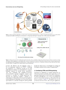
Figure 3. Difference between bioink and biomaterial ink. In bioink, cells are compulsory components of the printing formulation in the form of single
cells/coated cells/cell aggregates or combinations with materials. In biomaterial ink, cells are treated with the tissue construct only after bioprinting and
post-printing treatments. Reprinted from Biofabrication, 11, Groll J, Burdick JA, Cho DW, et al., A definition of bioinks and their distinction from bioma-
terial inks, 013001, Copyright (2018), with permission from IOP Publishing .
[90]
promote cell adhesion and cell migration owing to include the characteristics of the bioinks for printing and
the chemoattractant properties of CNTs . Following the bioink constructs for the desired TE applications .
[87]
[89]
the preparation of a composite hydrogel of GO and
polyacrylamide, Hyerim et al. investigated the composite 3. Combining CFNs and 3D bioprinting
hydrogel’s capacity for skeletal muscle regeneration.
They have established that graphene-incorporated Following the compilation of recently publications on
electroconductive hydrogels upregulate myogenic gene CFNs incorporated in printable gel for applications in TE
expression in myoblasts via cellular interactions with and tissue regeneration, we also outline the contribution
electrical and mechanical signals . However, it is of CFNs in printable gel in this review. The printable gel
[88]
challenging to develop robust nanocomposite hydrogels for used for TE has been classified as a biomaterial ink and
both bioprinting and cell seeding. Li et al. described that the bioink, as shown in Figure 3 . Biomaterial inks are 3D
[90]
specifications of a candidate hydrogel for printing should printed and sterilized before cell seeding for application.
Volume 9 Issue 1 (2023) 185 https://doi.org/10.18063/ijb.v9i1.635