
Page 197 - IJB-9-1
P. 197
International Journal of Bioprinting 3D bioprinting of tissue with carbon nanomaterials
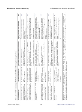
Ref. [104] [105] [106] [107] [108] [97]
Biological outcomes/ tissue engineering application An increase in oxygen metabolism and neural differentiation of the NSCs was observed at 25 ppm of GO or G-P. Neural tissue engineering SGOB1 bioink demonstrated larger electrocon- ductive and mechanical behavior and significant Neuro2a cell proliferation and cell viability com- pared to SB. Neural tissue engineering With GO in the gel, there was higher cell prolif- eration after 7
Bioprinters, crosslinking mechanisms, and scaffold’s
EBB (Regenovo 3D Bioprinter, China) Sol-gel thermal transition near 37°C Printed construct: 15 mm × 15 mm × 5 mm (L × W × H) PBB (Digital Light Processing Bioprinter, NBRTech Co., Ltd., Chuncheon South Korea) Methacrylation, carbodiimidation, and photoinitiation 3D square structure: 5 mm × 5 mm × 2 mm (L × W × H) EBB (IINVIVO, Rokit, Seoul, Korea) Enzymatic reaction by HRP and glucose oxidase Lattice cuboid 3D model: 2 cm × 2 cm × 0.2
Table 2. List of CFNs-containing cell-laden bioinks for different tissue engineering applications
dimensions by LAP × H) Photoinitiation by LAP Canada) 0.2 cm)
Cell-laden bioink composites PU, PU/GO 25 ppm, and PU/G-P 25 ppm NSCs (4 × 10 6 cells/mL) SB, 0.25% SGOB1, 2.5% SGOB1, 3.5% SGOB1, and 0.25% SGOB2 Neuro2a mouse neuroblastoma (1 × 10 7 cells/ mL) GHPA and GO-GHPA C2C12 skeletal myoblasts (1 or 5 × 10 5 cells/ mL) GelMA, GelMA/GO (GO: 0.02, 0.40, and 1.40 mg/mL) PC-12 rat pheochromocytoma cell line (5 × 10 4 cells/mL) MeCol and CNT-MeCol HCAECs (0.8–1 × 10 6 cells/mL) ABT,
CFNs and dimensions GO (Hummers’ method; LS = 4 µm) Multilayered pluronic-stabilized graphene, G-P (physical exfoliation method; LS = 2 µm) GO (obtained from Antopaar, S. Ko- rea, hydrodynamic diameter = 2.711 µm; BET surface area = 10.426 m 2 /g) GO (sheet diameter < 10 µm) (Sigma Aldrich, USA) GO (obtained from ANFF, Universi- ty of Wollongong, Australia; average diameter = 1–2 µm, single layer sheet with a thickness of 0.81 nm)
Volume 9 Issue 1 (2023) 189 https://doi.org/10.18063/ijb.v9i1.635