
Page 269 - IJB-9-1
P. 269
International Journal of Bioprinting Evaluation of advanced visual computing solutions for the left atrial appendage occlusion
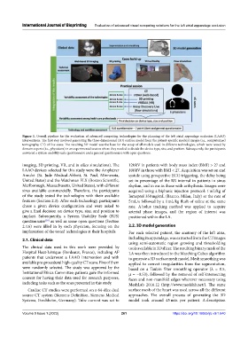
Figure 1. Overall pipeline for the evaluation of advanced computing technologies for the planning of the left atrial appendage occlusion (LAAO)
interventions. The first step involved generating the three-dimensional (3D) surface model from the patient-specific medical images (i.e., computerized
tomography, CT) of five cases. The resulting 3D model was the base for the setup of all models used in different technologies, which were tested by
domain experts (i.e., physicians) in an experimental session where they needed to decide the device type, size, and position. Subsequently, the participants
answered a system usability scale questionnaire and a general questionnaire with open questions.
imaging, 3D printing, VR, and in silico simulations). The 120 kV in patients with body mass index (BMI) > 27 and
LAAO devices selected for this study were the Amplatzer 100 kV in those with BMI < 27. Acquisition was set on end
Amulet (St. Jude Medical-Abbott, St. Paul, Minnesota, systole using prospective ECG triggering, the delay being
United States) and the Watchman FLX (Boston Scientific, set in percentage of the RR interval in patients in sinus
Marlborough, Massachusetts, United States), with different rhythm, and in ms in those with arrhythmia. Images were
sizes available commercially. Therefore, the participants acquired using a biphasic injection protocol: 1 mL/kg of
of the study tested the technologies with their available Iomeprol 350 mg/mL (Bracco, Milan, Italy) at the rate of
features (Section 2.3). After each technology, participants 5 mL/s followed by a 1 mL/kg flush of saline at the same
chose a given device configuration and were asked to rate. A bolus tracking method was applied to acquire
give a final decision on device type, size, and position to arterial phase images, and the region of interest was
implant. Subsequently, a System Usability Scale (SUS) positioned within the LA.
questionnaire as well as some open questions (Section
[38]
2.4.6) were filled in by each physician, focusing on the 2.2. 3D model generation
implantation of the tested technologies at their hospitals. For each selected patient, the anatomy of the left atria,
2.1. Clinical data including its appendage, was extracted from the CT images
using semi-automatic region growing and thresholding
The clinical data used in this work were provided by tools available in 3D slicer. The resulting binary mask of the
Hospital Haut-Lévêque (Bordeaux, France), including AF LA was then introduced to the Marching Cubes algorithm
patients that underwent a LAAO intervention and with to generate a 3D surface mesh model. Mesh smoothing was
available pre-procedural high-quality CT scans. Five of them applied to correct irregularities from the segmentation,
were randomly selected. The study was approved by the based on a Taubin filter smoothing operator (λ = 0.5,
Institutional Ethics Committee; patients gave the informed µ = −0.53), followed by the removal of self-intersecting
consent for having their data used for research purposes, faces and non-manifold edges wherever necessary using
including tasks such as the ones presented in this study. MeshLab 2016.12 (http://www.meshlab.net/). The same
Cardiac CT studies were performed on a 64-slice dual surface mesh of the heart was used across all the different
source CT system (Siemens Definition, Siemens Medical approaches. The overall process of generating the 3D
Systems, Forchheim, Germany). Tube current was set to model took around 45 min per patient. A description
Volume 9 Issue 1 (2023) 261 https://doi.org/10.18063/ijb.v9i1.640