
Page 273 - IJB-9-1
P. 273
International Journal of Bioprinting Evaluation of advanced visual computing solutions for the left atrial appendage occlusion
together with the full range of 3D-printed replicas of the A B
Amplatzer Amulet and Watchman FLX LAAO devices
available in the market. Physicians could then interact with
both types of 3D-printed models to decide which device
would fit each LA anatomy better.
2.4.4. Virtual reality VRIDAA platform
The tasks performed by the participants on the VRIDAA
platform were very similar to VIDAA. Nevertheless, the
LAA centerline was already provided by default. Once the
participant wore the VR glasses, the LA appeared, together C d
with its centerline, and the physician could move it, grab it,
or go inside to better explore the interior of the anatomical
structure. Then, the user could select the type and size of
a given device, which would be placed at the beginning of
the centerline, which was able to move along. In addition,
the LAAO device could also be grabbed and moved freely.
2.4.5. In silico simulations
Initially, the participant was asked to select a device
type and size, after which it could be freely placed in
any location of the LAA using the interface of the Ansys
Discovery Live software. The LA anatomy could also be
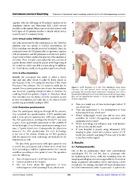
moved. Once a device position was chosen, the simulation Figure 5. (A‑D) Examples of in silico fluid simulations using Ansys
Discovery Live with optimal device settings according to a given
was launched, requiring several minutes to visualize the participant. 3D streamline-based visualization simulations were used to
resulting blood flow patterns (Figure 5). Therefore, blood illustrate blood flow patterns of in the left atrium and left atrial appendage.
flow velocities near the device could be estimated, as well Blue and red represent low velocity (< 10 m/s) and high velocities (>
as flow recirculations and leaks due to the chosen LAAO 20 m/s), respectively.
positioning, potentially leading to DRT.
• Have you tested any of these technologies before? If
2.4.6. Evaluation questionnaires yes, which one?
• Have you participated in the development of these
Once the participants had gone through all the patients, technologies? If yes, which one?
they answered two questionnaires: A SUS questionnaire • Which technologies would you add in your ideal
and a more general questionnaire with open questions. workflow for LAAO (disregarding economical and
The SUS questionnaire, developed by Brooke , was used equipment restrictions)?
[38]
to give a more quantitative assessment on the usability of • Which technology did influence your final decision
the technologies. It is a 10-item questionnaire using, in our on device election the most?
case, a 7-point Likert scale. Consequently, the physicians • If your hospital is mainly using ultrasound (US)
answered the SUS questionnaire for each technology imaging to plan LAAO interventions instead of CT,
at the end of the session. Details on the SUS questions would you consider acquiring CT data only to be able
and the answers for each technology are included in the to use these technologies?
Supplementary File.
The aim of the questionnaire with open questions was 3. Results
to profile the participants and to know more about how 3.1. Participant profile
these technologies could be implemented in their current Out of the six participants, three were interventional
workflow, according to their point of view. The questions cardiologists (P2, P3, and P4), that is, physicians who
were the following:
are implanting the device, while the remaining three are
• Years of experience in LAAO interventions. imaging cardiologists, who were responsible for the medical
• Current position at the hospital. image acquisition and analysis before and during LAAO
• Did you know about the application of these procedures. On average, they had 5.08 years of experience
technologies to LAAO planning? If yes, which one? in LAAO interventions (with 10 years and < 1 year for the
Volume 9 Issue 1 (2023) 265 https://doi.org/10.18063/ijb.v9i1.640