
Page 271 - IJB-9-1
P. 271
International Journal of Bioprinting Evaluation of advanced visual computing solutions for the left atrial appendage occlusion
approximately. The 3D LA model was prepared for A
printing with the Ultimaker Cura 3 software from the
printer provider. Typical rigid polylactic acid (PLA)
was the material used for the LA models (Figure 3),
with an associated cost of 1.5 euros for each model.
Twenty hours were needed to print all LA cases (i.e.,
4 h/case). Moreover, CAD models of Amplatzer and
Watchman FLX LAAO devices corresponding to the
different commercially available sizes were printed with
thermoplastic polyurethane (TPU) to add more flexibility. B
Therefore, users could try to position the printed LAAO
device into the 3D-printed model of the LA to have more
insight into their interaction. The cost of all printed
LAAO devices was of 10 euros, taking 22 h to print. An
extra hour was added for time estimations due to pre- and C
post-processing tasks, such as adding thickness to the 3D
models and removing the scaffolds of all the models.
2.3.3. VRIDAA: VR tool
The VRIDAA platform developed by Medina et al.
[27]
was used to evaluate the use of VR technologies for the
planning of LAAO interventions. VRIDAA allows the
user to interact with the LA geometry, jointly visualize
it with patient-specific medical images in standard MPR
format and relevant morphological indices (Figure 4).
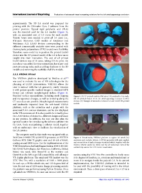
Standard surface manipulation, including mesh clipping Figure 3. (A) 3D-printed models of left atria (LA) analyzed in this study.
and transparency changes, as well as browsing along the (B) 3D-printed models of the left atrial appendage occluder (LAAO)
CT scan slices are possible. Morphological measurements devices. (C) Example of interaction between LA and LAAO 3D-printed
and landmarks imported from the web-based VIDAA models.
platform, such as the centerline and a graph with the
associated LAA contour diameters, can be also displayed
in the VR environment. It is also possible to virtually place
the LAAO device of choice (i.e., different designs and sizes)
in any position. In addition, the user can also plan the
optimal location for introducing the delivery catheter into
the LAA, freely manipulating a catheter model, together
with an endoscopic view to facilitate the visualization of
the LA interior.
The computer used in this study was equipped with an
Intel Core i5-8400 CPU @2.80 GHz processor, an NVIDIA Figure 4. Virtual reality VRIDAA platform to explore left atrial (LA)
GeForce RTX 2080 Ti graphic card and 16 GB of RAM, anatomies and occluder devices. A 3D LA geometry with axial, sagittal,
costing around 2000 euros. For the implementation of the and coronal slices of medical images visualized behind, together with the
VRIDAA platform, the Unity Engine version 2018.1.8f1 (64- delivery catheter model (in white) and the 2D endoscopic view of the
catheter’s tip camera for LAA interior visualization.
bit) (Unity Technologies, San Francisco, California, United
States) was used, with SteamVR as the runtime and
OpenVR as the API to get full compatibility with all major meshes using the HTC Vive Pro controller to freely move
VR display platforms. The employed VR headset was the it (6 degrees of freedom, i.e., rotations and translations) or
HTC Vive Pro, with a resolution of 1440 × 1600 pixels zoom it to navigate inside the patient’s LA. In its current
for each eye, a 90 Hz refresh rate, and 110 degrees field of implementation, the VRIDAA platform is intended to
view. Its cost was of 1239 euros. Image processing outside be used with the possibility of moving within the virtual
the platform was performed using Python libraries. Once environment. Thus, a clear space of around 2 m × 1.5 m is
uploaded into VRIDAA, the user can interact with the 3D required.
Volume 9 Issue 1 (2023) 263 https://doi.org/10.18063/ijb.v9i1.640