
Page 270 - IJB-9-1
P. 270
International Journal of Bioprinting Evaluation of advanced visual computing solutions for the left atrial appendage occlusion
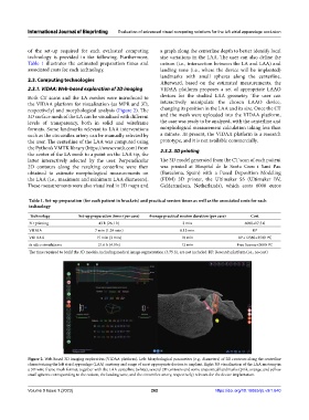
of the set-up required for each evaluated computing a graph along the centerline depth to better identify local
technology is provided in the following. Furthermore, size variations in the LAA. The user can also define the
Table 1 illustrates the estimated preparation times and ostium (i.e., intersection between the LA and LAA) and
associated costs for each technology. landing zone (i.e., where the device will be implanted)
landmarks with small spheres along the centerline.
2.3. Computing technologies
Afterward, based on the estimated measurements, the
2.3.1. VIDAA: Web-based exploration of 3D imaging VIDAA platform proposes a set of appropriate LAAO
Both CT scans and the LA meshes were introduced to devices for the studied LAA geometry. The user can
the VIDAA platform for visualization (as MPR and 3D, interactively manipulate the chosen LAAO device,
respectively) and morphological analysis (Figure 2). The changing its position in the LAA and its size. Once the CT
3D surface mesh of the LA can be visualized with different and the mesh were uploaded into the VIDAA platform,
levels of transparency, both in solid and wireframe the case was ready to be analyzed, with the centerline and
formats. Some landmarks relevant to LAA interventions morphological measurement calculation taking less than
such as the circumflex artery can be manually selected by a minute. At present, the VIDAA platform is a research
the user. The centerline of the LAA was computed using prototype, and it is not available commercially.
the Python’s VMTK library (https://www.vmtk.com) from 2.3.2. 3D printing
the center of the LA mesh to a point on the LAA tip, the
latter interactively selected by the user. Perpendicular The 3D model generated from the CT scan of each patient
2D contours along the resulting centerline were then was printed at Hospital de la Santa Creu i Sant Pau
obtained to estimate morphological measurements on (Barcelona, Spain) with a Fused Deposition Modeling
the LAA (i.e., maximum and minimum LAA diameters). (FDM) 3D printer, the Ultimaker S5 (Ultimaker BV,
These measurements were also visualized in 2D maps and Geldermalsen, Netherlands), which costs 6000 euros
Table 1. Set‑up preparation (for each patient in brackets) and practical session times as well as the associated costs for each
technology
Technology Set‑up preparation times (per case) Average practical session duration (per case) Cost
3D printing 43 h (26.1 h) 3 min 6000+17.5 €
VIDAA 7 min (1.24 min) 8.13 min RP
VRIDAA 15 min (3 min) 10 min RP+1238€+2500 PC
In silico simulations 21.6 h (4.3 h) 12 min Free license+2000 PC
The time required to build the 3D models, including medical image segmentation (3.75 h), are not included. RP: Research platform (i.e., no cost)
Figure 2. Web-based 3D imaging exploration (VIDAA platform). Left: Morphological parameters (e.g., diameters) of 2D contours along the centerline
characterizing the left atrial appendage (LAA) anatomy and range of most appropriate devices to implant. Right: 3D visualization of the LAA anatomy in
a 3D wire-frame mesh format, together with the LAA centerline (white), several 2D contours and some anatomical landmarks (pink, orange, and yellow
small spheres corresponding to the ostium, the landing zone, and the circumflex artery, respectively) relevant for the device implantation.
Volume 9 Issue 1 (2023) 262 https://doi.org/10.18063/ijb.v9i1.640