
Page 277 - IJB-9-1
P. 277
International Journal of Bioprinting Evaluation of advanced visual computing solutions for the left atrial appendage occlusion
comfortable using it. It can be observed in Table 4 that in 3D printing solutions are complementary, all providing
most cases, participants chose up to three different LAAO added value in different steps of the current LAAO clinical
device sizes, generally (63% of the times) not matching workflow. All the evaluated technologies passed the
the implanted one and with some extreme choices (C4 in threshold of acceptability range on usability; the web-based
Table 4, with A18 and A25 being selected). VIDAA platform and 3D printing were better rated, and
the former getting excellent marks from some participants.
4. Discussion The VR-based platform (VRIDAA) and in silico simulations
The fields of visual computing and 3D printing have seen were placed in the high and low marginal ranges,
a considerable progress over the last few years, slowly respectively, but huge discrepancies between participants
providing solutions for advanced visualization in some existed in the latter. However, we need to consider that
biomedical applications. According to a recent review previous experience on the technologies could influence
by Wang , a well-known cardiology leader in the field the usability score (e.g., P4 and P6 being the only users with
[2]
of LAAO interventions, 3D printing, computational some knowledge on simulations gave them the best scores).
modeling, and artificial intelligence (AI) plays a role in Nonetheless, the main overall conclusions among the
bridging the dichotomy of real-world in the trenches participants were the complementarity of the technologies
imaging and futuristic capabilities of computer science and and the need for an integrative unique platform of the
biomedical engineering. Pre-planning complex cardiology visual computing technologies (i.e., VIDAA, VRIDAA,
procedures such as LAAO with visual computing and 3D and fluid simulations) to be incorporated into the clinical
printing technologies can be beneficial in maximizing workflow and used on a daily basis. In addition, a more
interventional efficiency and minimizing costs, which realistic elastic behavior of the 3D-printed LAAO devices
are estimated by procedural time and device expenditure. would increase the precision on the selected settings.
However, the clinical translation of advanced visualization One of the most valued features in the web-based
technologies is not straightforward, needing to fulfill the VIDAA platform was the detailed characterization of the
demanding requirements to be embedded in the existing LAA anatomy, with the diameters along the centerline,
workflows in hospitals. For instance, clinicians will not since it can be used to identify the optimal implantation
invest more than a few minutes on planning cases that can or to better plan special strategies such as the sandwich
take 30 min for the intervention. Moreover, pre-planning technique. However, the manual selection of seed points
should not add excessive complexity and cost to the overall for the centerline was an important factor to properly
clinical workflow; therefore, sufficient cost-effectiveness characterize the LAA anatomy and select the appropriate
or patient safety impact must be proven to justify their device and its position. In addition, based on the SUS
routine use. questionnaire, VIDAA was fast and intuitive, which
Despite recent generic reviews of visual computing covered several manual steps but with a fast learning
solutions in cardiology applications [2,4-6] , there is a lack curve. Participants stressed the added value of the web-
of complete studies testing the different visualization based platform in complex cases, proposing to incorporate
methodologies on the same patient-specific data for functional information from fluid simulations for a more
benchmarking purposes. To the best of our knowledge, complete solution. The current commercial solutions
our study is the first attempt on this direction focusing on comparable to VIDAA are the stand-alone 3mensio
LAAO interventions, aiming at evaluating the added value, Structural Heart software (Pie Medical Imaging, Bilthoven,
limitations, and requirements for the clinical translation the Netherlands) or Mimics (Materialise NV, Leuven,
of these technologies. The results obtained in the practical Belgium), which include MPR visualization, 3D rendering,
session demonstrated that the tested visual computing and and 3D surface visualization, and HEARTguideTM
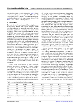
Table 4. Devices finally selected by the participants
Case P1 (I) P2 (IC) P3 (IC) P4 (IC) P5 (I) P6 (I) Implanted
C1 A22* A25 A22* A22* A28 A22* A22
C2 A18 A18 A22 A16* A22 A18 A16
C3 A20 A22 A22 A18* A22 A18* A18
C4 A20 W24 A20 A18 A22 A18 A25
C5 A22 A25* A25* A25* A25* W20 A25
W: Watchman flex; A: Amplatzer Amulet. Numbers refer to the device size (in mm). *The cases that matched the size of the device implanted to the
patient
Volume 9 Issue 1 (2023) 269 https://doi.org/10.18063/ijb.v9i1.640