
Page 169 - IJB-9-2
P. 169
International Journal of Bioprinting 3D Printing Multifunctional Orthopedic Biocoatings
Table 2. Experimental design and ink composition for vancomycin release measurements and antimicrobial studies
Sample code Polymer type Polymer concentration (%wt) Vancomycin concentration (% w/v) ACP concentration (% w/v) No. of layers
R-1 PCL 1 1 0 20
R-2 PCL 1 2 0 10
R-3 PCL 1 2 1 20
A B
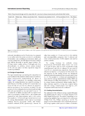
Figure 1. (A) Schematic of the custom 3D direct-write inkjet equipment. (B) Experimental setup for the deposition of composite polymeric formulations
and in situ infiltration.
polymeric formulations on the Ti substrates. As compared other three samples (n = 3) were used for in vitro viability
to our earlier work, this system consists of a combination and cytocompatibility assessment. Bare Ti substrate and
of deposition units including microextrusion, inkjet, and tissue culture polystyrene (TCPS) samples were used as
valve jet systems that can both deposit 3D tissue scaffolds positive controls.
and infiltrate biomedia at specific target locations. The The coating thickness for antibiotic release
UV/laser system enables curing of hydrogels and other measurements and bacterial culture studies was varied
photopolymers. In addition, camera system mounted at 10 and 20 layers, with the ACP concentration being
on the unit can capture real-time deposition images for varied between 0 and 1% w/v whereas VA was varied at
closed-loop feedback. 1 and 2% w/v concentrations, respectively. The polymeric
2.4. Design of experiment solution was fixed at 1 wt.% polymer in TFE solvent. The
run sequence for the coating process was determined
The experimental design and starting ink compositions to randomly and each experimental run was replicated 5 times
print the films for the osteoblast culture/assay and antibiotic (n = 5). Two samples (n = 2) from each experimental run
release measurements characterization are shown in were used for coating characterization studies (SEM and
Tables 1 and 2, respectively. For osteoblast culture/assay FTIR) whereas the other three samples (n = 3) were used
studies, the coating thickness was varied by printing 10 for release kinetics and bacterial culture studies. Bare Ti
and 20 layers of the films, and the ACP concentration was alloy substrates used for printing were weighed before
varied at 0.5% w/v and 1% w/v, whereas the polymeric and after coating. Coated samples were kept in a 4°C
solution was fixed at 1 wt. % polymer in solvent. The run refrigerator until release study was conducted.
sequence for the coating process was determined randomly
and each experimental run was replicated 5 times (n = 5) 2.5. Coating characterization
to enable the variability associated with the experimental The uniformity of the different coating samples fabricated
units to be estimated. A total of 40 (n = 40) samples were was studied using the optical microscopy (Keyence VHX
prepared for both characterization and in vitro studies. 600K Digital Microscope). The surface morphology of the
Two samples (n = 2) from each experimental run were used coatings was studied using a scanning electron microscope
for coating characterization studies (optical microscopy, (Philips-XL30 FEG, Philips) operating at 10.0 kV. The
scanning electron microscopy [SEM], and Fourier- samples used for SEM analysis were coated with palladium
transform infrared [FTIR] spectroscopy), whereas the (Pd) using a sputter coater system to obtain a conductive
Volume 9 Issue 2 (2023) 161 https://doi.org/10.18063/ijb.v9i2.661