
Page 172 - IJB-9-2
P. 172
International Journal of Bioprinting 3D Printing Multifunctional Orthopedic Biocoatings
This can be attributed to the precipitation and saturation processes deliver high-quality structures, however, need
of the ACP within the coated regions. further post-processing to achieve the desired functional
On the contrary, the PCL-ACP-coated substrates have a properties which can be overcome by employing the
uniform deposition (Figure 4A and 4B) pattern as seen under customized inkjet process implemented in this research.
the optical microscope, which is desirable for orthopedic 3.3. Chemical composition analysis
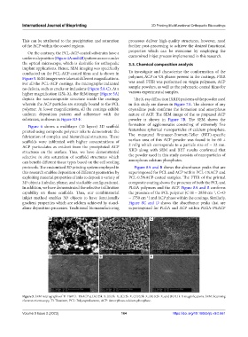
implant applications. Hence, SEM imaging was specifically
conducted on the PCL-ACP-coated films and is shown in To investigate and characterize the conformation of the
Figure 5. SEM images were taken at different magnifications. polymer, ACP or VA phases present in the coatings, FTIR
For all the PCL-ACP coatings, the micrographs indicated was used. FTIR was performed on virgin polymers, ACP
no defects, such as cracks or inclusions (Figure 5A-C). At a sample powders, as well as the polymeric-coated films for
higher magnification (25k-X), the SEM image (Figure 5A) various experimental samples.
depicts the nanocomposite structure inside the coatings The X-ray diffraction (XRD) patterns of the powder used
wherein the ACP particles are strongly bound to the PCL in this study are shown in Figure 7A. The absence of any
polymer. At lower magnifications, all the coatings exhibit crystalline peak confirms the formation and amorphous
uniform deposition pattern and adherence with the nature of ACP. The SEM image of the as prepared ACP
substrates, as shown in Figure 5D-F. powder is shown in Figure 7B. The SEM shows the
Figure 6 shows a multilayer (10 layers) 3D scaffold formation of agglomerates consisting of extremely fine
printed using composite polymer inks to demonstrate the featureless spherical nanoparticles of calcium phosphate.
fabrication of complex and hierarchical structures. These The measured Brunauer-Emmett-Teller (BET)-specific
scaffolds were infiltrated with higher concentrations of surface area of this ACP powder was found to be 60 ±
2
ACP particulates as evident from the precipitated ACP 2 m /g which corresponds to a particle size of ~ 32 nm.
structures on the surface. Thus, we have demonstrated XRD along with SEM and BET results confirmed that
selective in situ saturation of scaffold structures which the powder used in this study consists of nanoparticles of
can benefit different tissue types based on the cell seeding amorphous calcium phosphates.
protocols. The customized 3D printing system employed in Figure 8A and B shows the absorbance peaks that are
this research enables deposition of different geometries by superimposed for PCL and ACP within PCL-1%ACP and
exploiting material properties of inks to deposit a variety of PCL-0.5%ACP-coated samples. The FTIR of the printed
3D objects (tubular, planar, and stackable configurations). composite coating shows the presence of both the PCL and
In addition, we have demonstrated the selective infiltration PLGA polymers and the ACP. Figure 8A and B confirms
capability on these scaffolds. Thus, our combinatorial the presence of the PCL polymer (C-H ~ 2850 cm , C=O
−1
inkjet method enables 3D objects to have functionally ~ 1750 cm ) and ACP phase within the coatings. Similarly,
−1
gradient properties which are seldom achieved by stand- Figure 8C and D shows the absorbance peaks that are
alone deposition processes. Traditional biomanufacturing superimposed for PLGA and ACP within PLGA-1%ACP
A B C
D E F
Figure 5. SEM micrographs of Ti-1%PCL-1%ACP at (A) 25k-X, (B) 5k-X, (C) 2k-X, (D) 0.5k-X, (E) 0.2k-X, and (F) 0.1k-X magnifications. SEM: Scanning
electron microscopy, Ti: Titanium, PCL: Polycaprolactone, ACP: Amorphous calcium phosphate.
Volume 9 Issue 2 (2023) 164 https://doi.org/10.18063/ijb.v9i2.661