
Page 177 - IJB-9-2
P. 177
International Journal of Bioprinting 3D Printing Multifunctional Orthopedic Biocoatings
The fast degradation of PCL in contact with the buffer or and net charge [85,88] . Thus, the adsorption of VA molecules
presence of free VA particles on the film surface due to on the surfaces of nanosized ACP particles is highly
the poor encapsulation of VA inside the PCL films most feasible. On contact with water, some of these adsorbed
likely caused the burst release of VA at the beginning of VA molecules diffused out in the solution, thereby
elution for samples R-1 and R-2. However, the degradation resulting in a more sustained release in sample R-3. The
of PCL under physiological buffers has shown to be very decrease in the rate of release of VA over time is presumed
slow, and therefore, rapid dissolution of the inadequately partially due to the reduction in easily soluble amorphous
encapsulated VA causes the burst release . Moreover, content of the ACP powder on the particle surface, combined
[81]
this rapid dissolution of the VA should create pores within with the conversion of the ACP phase into hydroxyapatite
the PCL matrix. In the latter time points, the rest of the by dissolution precipitation as well as the strongly adsorbed
VA molecules and water molecules diffuse through these drug molecules on the particle surface [85,89] .
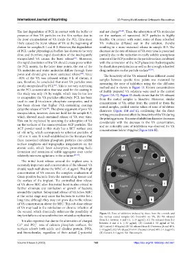
pores and should give a more sustained release . Since The bioactivity of the VA released from different coated
[82]
~80% of the VA was released within 8 h of elution, it samples between specific time points was measured by
can, therefore, be concluded that most VA particles were measuring the zone of inhibition using the disc diffusion
poorly encapsulated by PCL . This is not very surprising method and is shown in Figure 13. Known concentrations
[83]
as the PCL concentration that was used for the coating in of freshly prepared VA solutions were used as the control
this study was only 1% by weight, which may be too low (Figure 13A-H). Figure 13 clearly shows that the VA released
to encapsulate the VA particles effectively. PCL has been from the coated samples is bioactive. Moreover, similar
used to coat β-tricalcium phosphate composites, and it concentration of VA, either from the control or from the
has been shown that higher PCL-containing coatings coated samples, yielded similar values of zone of inhibition
delay the release of VA . The above argument of poor VA diameters (Figure 13B and E), confirming that the direct
[84]
encapsulation, however, does not hold for the sample R-3, writing process does not affect the bioactivity of the VA during
which showed much-sustained release of VA over time. the printing process. The zone of inhibition diameter decreases
This can be explained by assuming the adsorption of VA considerably with the decrease in the VA concentration,
on the surfaces of the nanocrystalline ACP particles. The and no noticeable zone of inhibitions was observed for VA
ACP powder used in this study has a BET surface area concentrations below 10 µg/ml (Figure 13A-H).
of ~61 m /g, which corresponds to spherical particles of
2
~32 nm in size. It is well-established in the literature that
these nanosized calcium phosphate particles also exhibit
surface roughness and topographic irregularities on the
atomic scale, which favor adsorption, promoting facile
formation and retention of stable aggregates even under
relatively intensive agitations in the solution [85-87] .
The initial burst release around the implant area is
extremely important and concentration of the released VA
should reach well above the MBC of ~8 µg/ml. This high
concentration of VA ensures the complete eradication of
Gram-positive bacteria from the surrounding tissues and
the surface of the implant. The controlled slow release
of VA above MIC after this initial burst is also critical to
further eliminate any reinfection or growth of bacteria
around the implant. Suboptimal release of VA below MBC
at the initial stage may cause the bacteria to survive for a
long time although they may not grow due to the release
of VA concentration above the MIC. This sub-dose release
of VA may lead to the reinfection or chronic infection of
the wound, which drastically enhances the possibility of Figure 13. Zone of inhibition induced by elutes from the controls and
implant failure and wound infection-related complications. the various coated samples (A) Control-1: no VA, (B) VA released
It is also reported that due to the alternation of charged from R-1 between 0 and 4 h. (~37 µg/ml), (C) VA released from R-2
Ca and PO ions of calcium phosphate surfaces, the between 0 and 4 h. (~32 µg/ml), (D) Control-2: 50 µg/ml of VA,
3−
2+
(E) Control-3: 40 µg/ml, (F) VA released from R-3 between 24 and 48 h.
4
surfaces adsorb both acidic and alkaline protein, DNA, (~10 µg/ml), (G) VA released from R-2 between 24 and 48 h. (~1.6 µg/ml),
and biomolecules, regardless of their actual ζ-potential (H) Control-3: 5 µg/ml. VA: Vancomycin.
Volume 9 Issue 2 (2023) 169 https://doi.org/10.18063/ijb.v9i2.661