
Page 174 - IJB-9-2
P. 174
International Journal of Bioprinting 3D Printing Multifunctional Orthopedic Biocoatings
A B
C D
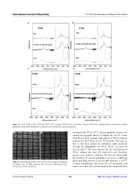
Figure 8. (A-D) FTIR of PCL-ACP and PLGA-ACP coatings. FTIR: Fourier transform infrared, PCL-ACP: Polycaprolactone-amorphous calcium
phosphate, PLGA-ACP: Poly(lactic-co-glycolic) acid-amorphous calcium phosphate.
A B correlated the PLGA-ACP cytocompatibility results with
optical micrographs shown in Figure 4C and D, which
show PLGA-ACP coatings with regions of PLGA polymer
without the ACP phase on the Ti substrate. This may be
due to the local release of carboxylic acids produced
through the degradation of PLGA, which increases the
local acidity . We have shown that the degradation of
[69]
PLGA reduces the local pH drastically and, therefore,
creates a zone which is cytotoxic. We have also shown that
the presence of calcium phosphate can act as a buffering
agent and help prevent a considerable decrease in pH .
[69]
Figure 9. Optical images of PCL-ACP_0.5% 10-layer coating on Ti substrate
(A) before and (B) after adhesion test. PCL-ACP: Polycaprolactone- A more physiological pH favors the cell attachment and
amorphous calcium phosphate. hence, more live cells can be found in the composite films
Volume 9 Issue 2 (2023) 166 https://doi.org/10.18063/ijb.v9i2.661