
Page 175 - IJB-9-2
P. 175
International Journal of Bioprinting 3D Printing Multifunctional Orthopedic Biocoatings
of PLGA-ACP. Except for the very few of these localized The presence of ACP in the printed films, however,
depositions of the PLGA patches, the rest of the film allows offers some unique advantages other than buffering
the cells to attach and proliferate. The high % viability of the local pH. It is well-known that ACP has the highest
these PLGA-ACP films (Figure 10) clearly demonstrates solubility among the various calcium phosphate phases,
that these PLGA-rich zones have hardly any effect on the and therefore, it is expected to dissolve and release calcium
overall cell viability and proliferation. and phosphate ions in the system . Moreover, the
[70]
protons generated from the released acidic byproducts of
PCL and PLGA interact with the ACP particles, leading
to an increase in dissolution of the ACP particles which
also causes an increase in the concentrations of soluble
Ca and phosphate in the surrounding media. It is well-
2+
established that the release of calcium and phosphate ions
locally improves the osteoclast and osteoblast activity,
which, in turn, facilitates bone regeneration [71,72] . Thus, it is
expected that the composite films of PCL-ACP and PLGA-
ACP should demonstrate improved biological response as
compared to the polymers film alone.
3.7. Antibiotic drug release kinetics
The in vitro release kinetics of the VA from the coated
samples R-1, R-2, and R-3 samples (Table 2) was measured
in PBS and is shown in Figure 12A. The samples R-1
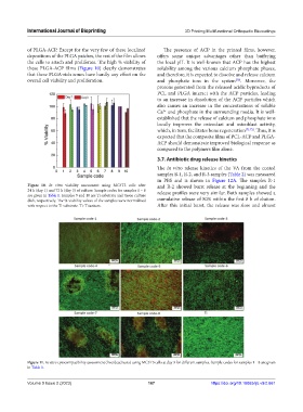
Figure 10. In vitro viability assessment using MC3T3 cells after and R-2 showed burst release at the beginning and the
24 h (day 1) and 72 h (day 3) of culture. Sample codes for samples 1 – 8 release profiles were very similar. Both samples showed a
are given in Table 1. Samples 9 and 10 are Ti substrate and tissue culture
dish, respectively. The % viability values of the samples were normalized cumulative release of 80% within the first 8 h of elution.
with respect to the Ti substrate. Ti: Titanium. After this initial burst, the release was slow and almost
Figure 11. In vitro cytocompatibility assessment (live/dead tests) using MC3T3 cells at day 3 for different samples. Sample codes for samples 1 – 8 are given
in Table 1.
Volume 9 Issue 2 (2023) 167 https://doi.org/10.18063/ijb.v9i2.661