
Page 304 - IJB-9-2
P. 304
International Journal of Bioprinting Zn-doped coatings with osteogenic and antibacterial properties
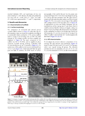
standard deviation (SD), and significance of data was micrographs of HA scaffold showed that the grains were
determined by P < 0.05. Besides, the data with probability compacted (Figure 3D).”. As for the morphology of coating,
less than 0.05 (P < 0.05), 0.01 (P < 0.01), and 0.001 the coating materials penetrated into the gaps between
(P < 0.001) were represented by *, **, and ***, respectively. ceramic particles and a smoother surface was then obtained
(Figure 3E). The cross-section morphology of the scaffold
3. Results and discussion with coatings is shown at the top right corner of Figure 3E.
3.1. Characterization of scaffolds A multilayered structure was clearly displayed, and the
total thickness of the coating was 6.7 μm, from which it
3.1.1. Surface morphology could be inferred that the thickness of single coating was
The comparison of unsintered and sintered porous about 0.74 μm. In addition, the change of Zn concentration
ceramic scaffold is shown in Figure 3A. After sintering, the in the coating had no effect on its morphology. Finally, the
photosensitive resin was removed completely, resulting in EDS mapping of Ca and Zn elements in the coating of the
a considerable volume contraction of the porous scaffold, CHA-H scaffold confirmed that Zn was homogeneously
2+
and the average linear shrinkage rate was 30%. The XRD distributed (Figure 3G and 3H).
patterns of the ceramic powder and bare scaffolds are
displayed in Figure 3B. The main component of the 3.1.2. XPS characterization
scaffold was HA (# 70-0566), and no significant phase To investigate the detailed chemical composition of the
transition occurred during sintering. Compared with coatings, the XPS analyses were performed. Figure 4A
the size distribution of raw HA powders (Figure 3C), the shows the total XPS spectra of CHA-0 and CHA-H groups,
particle size of HA scaffold surface mainly distributed from which were typical among all groups. The elements
10 to 15 μm (Figure 3D), which indicated that the grain contained in the raw material, such as C, N, O, Ca, and
grew normally. In addition, the SEM results of surface Zn, could be detected in CHA-H, and all of these elements
A D F
B
G
E
C
H
Figure 3. (A) Images of unsintered and sintered hydroxyapatite (HA) scaffolds. (B) X-ray diffraction spectra of HA powder and scaffold. (C) Particle size
distribution of ceramic powders. (D) Scanning electron microscope (SEM) image of HA scaffold; the inset image shows the size distribution of ceramic
particle on the scaffold surface. (E) SEM image of HA scaffold with coatings; the inset image shows the cross section of coatings. (F) High-magnification
SEM image of coatings. (G and H) EDS mapping of Ca and Zn on CHA-H sample.
Volume 9 Issue 2 (2023) 296 https://doi.org/10.18063/ijb.v9i2.668