
Page 302 - IJB-9-2
P. 302
International Journal of Bioprinting Zn-doped coatings with osteogenic and antibacterial properties
water to remove the uncrosslinked solution and then 2.3. Characterization of scaffolds
placed in a conventional oven at 60°C for 2 min. Then, the The scanning electron microscope (SEM, SU5000, Hitachi
above-mentioned soaking process was repeated until all Instruments, Japan) was used to analyze the surface and
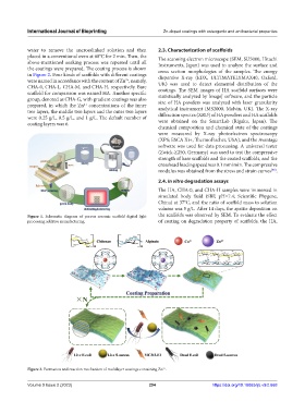
the coatings were prepared. The coating process is shown cross section morphologies of the samples. The energy
in Figure 2. Four kinds of scaffolds with different coatings dispersive X-ray (EDX, ULTIMATELYMAX40, Oxford,
were named in accordance with the content of Zn , namely, UK) was used to detect elemental distribution of the
2+
CHA-0, CHA-L, CHA-M, and CHA-H, respectively. Bare coatings. The SEM images of HA scaffold surfaces were
scaffold for comparison was named HA. Another specific statistically analyzed by ImageJ software, and the particle
group, denoted as CHA-G, with gradient coatings was also size of HA powders was analyzed with laser granularity
prepared, in which the Zn concentrations of the inner analytical instrument (MS2000, Malvin, UK). The X-ray
2+
two layers, the middle two layers and the outer two layers diffraction spectra (XRD) of HA powders and HA scaffolds
were 0.25 g/L, 0.5 g/L, and 1 g/L. The default number of were obtained on the SmartLab (Rigaku, Japan). The
coating layers was 6.
chemical composition and chemical state of the coatings
were measured by X-ray photoelectron spectroscopy
(XPS; ESCA Xi+, ThermoFischer, USA), and the Avantage
software was used for data processing. A universal tester
(Zwick-Z250, Germany) was used to test the compressive
strength of bare scaffolds and the coated scaffolds, and the
crosshead loading speed was 0.1 mm/min. The compressive
modulus was obtained from the stress and strain curves .
[33]
2.4. In vitro degradation assays
The HA, CHA-0, and CHA-H samples were immersed in
simulated body fluid (SBF, pH=7.4; Scientific Phygene,
China) at 37°C, and the ratio of scaffold mass to solution
volume was 5 g/L. After 14 days, the apatite deposition on
Figure 1. Schematic diagram of porous ceramic scaffold digital light the scaffolds was observed by SEM. To evaluate the effect
processing additive manufacturing. of coating on degradation property of scaffolds, the HA,
Figure 2. Formation and reaction mechanism of multilayer coatings containing Zn .
2+
Volume 9 Issue 2 (2023) 294 https://doi.org/10.18063/ijb.v9i2.668