
Page 307 - IJB-9-2
P. 307
International Journal of Bioprinting Zn-doped coatings with osteogenic and antibacterial properties
A C E
B D F
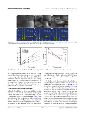
Figure 6. Scanning electron microscope images of mineral deposited on (A) hydroxyapatite (HA), (C) CHA-0, and (E) CHA-H scaffolds in SBF for 14 days.
EDS analyses of the surface sediment of (B) HA, (D) CHA-0, and (F) CHA-H scaffolds are also shown.
A B
Figure 7. Weight loss (A) and pH value (B) of scaffolds after soaking in Tris-HCl for different durations (the solution refreshed every week).
during the preparation of the coating. Although the pH capacities in decreasing order were: CHA-M, CHA-L, HA,
value of the solution after soaking the bare HA scaffold and CHA-0 groups. The low cell activity of CHA-H group
was on average higher than that of coated scaffolds, these was caused by high concentration of Zn ion, which was
2+
differences were not significant. Overall, the coating toxic to cells.
had little effect on the pH of body fluid environment. In To further verify biocompatibility of scaffolds, the
particular, the pH values of all groups were always higher cell physiological status and survival was observed using
than 7, showing weak alkalinity, which has been proven to fluorescence microscope on day 4. In Figure 9A, the
be beneficial to the growth of osteoblasts . cytoskeleton was displayed as green spots and the nucleus
[46]
3.4. In vitro biocompatibility properties was shown as blue spots. The filopodia and pseudopodia
extension of the cell could be conformably observed on
Although the addition of the coating inhibited apatite all scaffolds except CHA-H, which indicate excellent
formation, it was not the sole criterion for biological growth of cells . The cells on CHA-H scaffold surface
[47]
activity. The viability of MC3T3-E1 cells on days 1, 4, and were smaller and not extended enough. Furthermore,
7 was evaluated by CCK-8 assay. As shown in Figure 8, the the quantity of cells in each group shown by fluorescence
cell proliferation of all groups (except CHA-H) was better images was basically in accordance with the data of CCK-8
than that of blank control group in a time-dependent assay. To explore the damage on cells caused by high Zn
2+
manner, indicating good biocompatibility of the scaffolds. concentration, calcein and propidium iodide staining was
Furthermore, at each detection node, their proliferative performed. As shown in Figure 9B, living cells were green
Volume 9 Issue 2 (2023) 299 https://doi.org/10.18063/ijb.v9i2.668