
Page 393 - IJB-9-2
P. 393
International Journal of Bioprinting In situ 3D bioprinter for skin wound healing
In vivo experiments were performed at the Veterinary glasses, and routinely stained with hematoxylin-eosin dye
Department of the National Medical Research Center (BioVitrum, Russia).
for Radiology and were approved by the local ethics Morphometry histological sections were performed
committee (protocol #0120/19 dated November 1, 2019). using automated image analysis system (ImageJ, USA)
Throughout the experiment, the animals were kept in and quantitative morphometric parameters such as
individual ventilated boxes under exhaust ventilation and inflammatory index (number of inflammatory cells per
were fed ad libitum. The animal work was carried out in unit of analyzed histological section area) and angiogenesis
accordance with the ethical principles established by the index (number of microvessels profile per unit of analyzed
European Convention for the Protection of Vertebrate histological section area) were estimated with sequential
Animals used for Experimental and Other Scientific statistical analysis.
Purposes (Strasbourg, 2006) and the International
Guidelines for Biomedical Research in Animals (CIOMS 2.15. Statistical analysis
and ICLAS, 2012). Circular full-thickness skin defects were Statistical data were analyzed and graphs were plotted
formed using sterile surgical instruments after preliminary using GraphPad Prism software (GraphPad Software, Inc.,
intravenous Zoletil/Xylazine sedation of animals. In the La Jolla, CA) and expressed in mean ± standard deviation.
case of minipigs, the isoflurane inhalation anesthesia The Mann–Whitney U-test was used to compare the
was additionally used. Robotic-assisted bioprinting with quantitative characteristics of the groups. Statistical
hydrogel compositions was carried out immediately after significance was determined at P < 0.05.
the wound preparation and cleaning. The polymerized
bioprinted collagen hydrogels in wounds were carefully 3. Results
covered with dressings. All animals received intramuscular
antimicrobial and analgesic drugs for 1 week after surgery. 3.1. Feedback system
Since the patient is breathing during bioprinting, it is
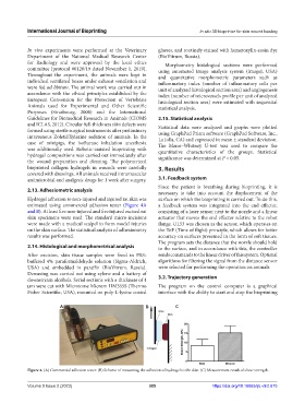
2.13. Adhesiometric analysis
necessary to take into account the displacement of the
Hydrogel adhesion to non-injured and injured rat skin was surface on which the bioprinting is carried out. To do this,
estimated using commercial adhesion tester (Figure 4A a feedback system was integrated into the end effector,
and B). At least five non-injured and five injured excised rat consisting of a laser sensor next to the nozzle and a linear
skin specimens were used. The standard micro-incisions actuator that moves the end effector relative to the robot
were made with a medical scalpel to form model injuries flange. UL53 was chosen as the sensor, which operates on
on the skin surface. The statistical analysis of adhesiometry the ToF (Time of flight) principle, which allows for better
results was performed. accuracy on surfaces presented in the form of soft tissues.
The program sets the distance that the nozzle should hold
2.14. Histological and morphometrical analysis to the surface, and in accordance with this, the controller
After excision, skin tissue samples were fixed in PBS- sends commands to the linear driver of this system. Optimal
buffered 4% paraformaldehyde solution (Sigma-Aldrich, algorithms for filtering the signal from the distance sensor
USA) and embedded in paraffin (BioVitrum, Russia). were selected for performing the operation on animals.
Dewaxing was carried out using xylene and a battery of
downstream alcohols. Serial sections with a thickness of 4 3.2. Trajectory generation
μm were cut with Microtome Microm HM355S (Thermo The program on the control computer is a graphical
Fisher Scientific, USA), mounted on poly-L-lysine coated interface with the ability to start and stop the bioprinting
A B C
Figure 4. (A) Commercial adhesion tester. (B) Scheme of measuring the adhesion of hydrogel to the skin. (C) Measurement result of shear strength.
Volume 9 Issue 2 (2023) 385 https://doi.org/10.18063/ijb.v9i2.675