
Page 395 - IJB-9-2
P. 395
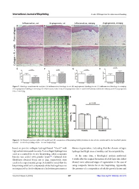
International Journal of Bioprinting                               In situ 3D bioprinter for skin wound healing


               A                       B                      C                       D


               E                       F                       G                      H


            Figure 5. Histology morphometric analysis: (A) inflammatory histology in rat; (B) angiogenesis histology in rat; (C) inflammatory histology in minipig;
            (D) angiogenesis histology in minipig; (E) inflammatory index in rat; (F) angiogenesis index in rat; (G) inflammatory index in minipig; and (H) angiogenesis
            index in minipig.

                         A                                   B


            Figure 6. (A) Printed construct with CAD model and (B) comparison of bioprinting fidelity between in situ robotic printer and in situ handheld printer
            (shaded – in vitro bioprinting, white – in vivo bioprinting).

            based on porcine collagen hydrogel brand “Viscoll” with   fibrous degeneration, indicating that the chosen collagen
            high adhesiveness and viscosity. Pure collagen hydrogel was   hydrogel had high rates of sterility and biocompatibility.
            used as a control for in situ bioprinting, while composite
                                            [33]
            bioinks was added with platelet lysate . Cultured skin   At the same time, a histological analysis performed
            fibroblasts  obtained  from  rats  or  pigs,  respectively,  were   4 weeks after the surgical formation of a full-layer skin defect
            used in the experimental group. It should be noted that the   showed more advanced stages of regeneration in the case of
            bioprinting with both compounds of the hydrogel was not   using composite bioinks for in situ bioprinting. Apparently,
            accompanied by the development of infectious processes or   the presence of a composition of soluble growth factors and


            Volume 9 Issue 2 (2023)                        387                      https://doi.org/10.18063/ijb.v9i2.675