
Page 120 - IJB-9-3
P. 120
International Journal of Bioprinting OMT-loaded spinal cord scaffold
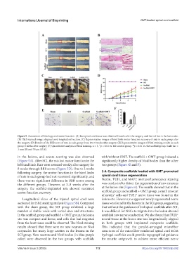
Figure 5. Assessment of histology and motor function. (A) Rat spinal cord tissue was obtained 8 weeks after the surgery, and the red box is the lesion site.
(B) H&E-stained image of spinal cord longitudinal section. (C) Representative images of hind limb motor function recovery of rats in each group after
the surgery. (D) Results of the BBB score of rats in each group from 1 to 8 weeks after surgery. (E) Representative images of Nissl staining results in each
group 8 weeks after surgery. (F) Quantitative analysis of Nissl staining. n = 3, p < 0.01 vs. the control group. p < 0.01 vs. the scaffold group. Scale bar =
**
##
1 mm (B) and 50 μm (B, E).
in the lesions, and severe scarring was also observed with/without OMT. The scaffold + OMT group induced a
(Figure 5A). After SCI, the rats lost motor function in the significantly higher density of Nissl bodies than the other
left hind limb. Rats were assessed weekly after surgery for two groups (Figure 5E and F).
8 weeks through BBB scores (Figure 5D). One to 2 weeks
following surgery, the motor function in the hind limbs 3.4. Composite scaffolds loaded with OMT promoted
of rats in each group had not recovered significantly, and spinal cord tissue regeneration
there was no significant difference in BBB scores among Nestin, TUJ1, and MAP2 immunofluorescence staining
the different groups. However, at 3–8 weeks after the was conducted to detect the regeneration of new neurons
surgery, the scaffold-implanted rats showed sustained at the lesion site (Figure 6). The results showed that in the
motor function recovery. scaffold group and scaffold + OMT group, a small amount
of nestin cells and TUJ1 nerve tissue was found in the
+
+
Longitudinal slices of the injured spinal cord were lesion site. However, no apparent newly regenerated nerve
sectioned for H&E staining analysis (Figure 5B). Compared tissue was found in the lesions in the SCI group, suggesting
with the sham group, the SCI group exhibited a large that without the guidance of biological scaffolds after SCI,
number of visible voids with varied sizes and structures. it was difficult for NSCs to migrate into the lesion site and
In the scaffold group and scaffold + OMT group, the lesion establish new nerve conduction. We also found that TUJ1
+
site was compact and dense, and cells that had migrated neural tissue at the lesion site was longitudinally aligned
from the host tissue could be observed. The Nissl staining in both groups with implanted composite scaffolds.
results showed that there were no new neurons or Nissl This indicated that the parallel-arranged microfiber
corpuscles but many large cavities in the lesions in the structures of the microfiber-reinforced spinal cord ECM
SCI group. New neurons and Nissl bodies (in blue-purple hydrogel scaffolds could provide topographical guidance
color) were observed in the two groups with scaffolds for neurite outgrowth to achieve more efficient nerve
Volume 9 Issue 3 (2023) 112 https://doi.org/10.18063/ijb.692