
Page 122 - IJB-9-3
P. 122
International Journal of Bioprinting OMT-loaded spinal cord scaffold
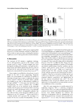
Figure 7. The composite scaffold alleviates the formation of glial scars. (A–D) Representative images of immunofluorescence in each group 8 weeks after
surgery. (A–A2) Normal spinal cord tissue in the sham group. (B–D, B1–D1) Compared with the SCI group, a small number of astrocytes were expressed
in the spinal cord lesions of the scaffold group and the scaffold + OMT group. (B2–D2) Compared with the SCI group, the expression of GFAP-positive
cells around the spinal cord lesions in the scaffold group and the scaffold + OMT group was significantly decreased; the scaffold + OMT group was more
significant. (E) GFAP immunofluorescence semi-quantitative analysis. (F) MAP2 immunofluorescence semi-quantitative analysis. n = 3, p < 0.01 vs. the
**
control group, p < 0.05 vs. the scaffold group. Scale bars = 1 mm (A–D) and 50 μm (A1–D1, A2–D2).
#
scaffold group and scaffold + OMT group, suggesting that low immunogenicity . Studies have found that spinal cord
[46]
the presence of OMT effectively inhibited the proliferation ECM-based hydrogels can regulate the behavior of NSCs and
and activation of astrocytes, hence reducing the formation promote their differentiation toward a neuronal lineage [31,47] .
of glial scars and eliminating obstacles to neurite extension. Our results showed that the composite scaffolds had excellent
biocompatibility, degradability, and mechanical property.
4. Discussion They could provide a stable and suitable microenvironment
for nerve regeneration and guide new axons to grow into
The treatment of SCI remains a significant challenge. the lesion site. These axons grew along with the longitudinal
As the self-regeneration capacity of the human central direction of the PCL microfiber bundles. Zhang et al. also
nervous system is weak , several strategies have been used electrospinning technology to prepare PCL/polysialic
[38]
developed, including cell implantation, hydrogel injection, acid hybrid multifunctional nanofiber scaffolds, and the
drug therapy, and scaffold implantation. However, effective scaffolds could promote axonal growth and functional
interventions for acute and chronic SCI are still limited, recovery after SCI in rats . Wang et al. constructed a
[48]
despite achievements in both basic and clinical research [38,39] . cytokine-loaded PCL–PEG composite hydrogel scaffold,
which could also guide the directional growth of axons .
[49]
Tissue engineering scaffolds have always been a research
hotspot in the field of tissue engineering. Numerous In addition, Xing et al. constructed an acellular spinal cord
scaffold cross-linked NT-3 sustained-release system, which
studies have demonstrated the advantages of scaffolds in promoted adhesion, proliferation, and differentiation of
delivering cells and drugs to repair or regenerate the central rat bone marrow mesenchymal stem cells . The slow
[50]
nervous system . However, in previous studies, scaffolds degradation of PCL can provide sufficient time for nerve
[38]
were mainly in the form of fibrous/porous scaffolds or regeneration for the growth of new axons at both ends and
hydrogel scaffolds [40-42] . In this study, near-field direct write the establishment of new connections.
electrospinning was used to fabricate PCL microfiber
bundles, which act as the backbone of the composite After SCI, axon regeneration is usually promoted by
scaffold, while the spinal cord ECM-based OMT-containing implanting exogenous NSCs or activating endogenous
hydrogel was used as the hydrogel matrix of the composite NSCs. Because NSCs have multi-directional differentiation
scaffolds. PCL, a synthetic biodegradable polyester, has potential, it is crucial to induce their differentiation toward
been fabricated into various biological scaffolds for nerve neurons. Some studies stimulated the activation and
regeneration and SCI research [43-45] . Spinal cord ECM is migration of endogenous NSCs by administering some
a natural material, and it possesses the advantages of a small biological/chemical molecules that promoted axon
natural 3D structure, excellent biocompatibility, and very regeneration [51,52] . In our study, the immunofluorescence
Volume 9 Issue 3 (2023) 114 https://doi.org/10.18063/ijb.692