
Page 211 - IJB-9-3
P. 211
International Journal of Bioprinting Dual ions mixed GelMA for hair follicle regeneration
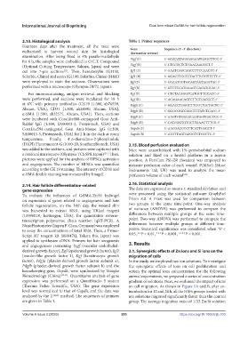
2.13. Histological analysis Table 1. Primer sequences
Fourteen days after the treatment, all the mice were Gene Sequence (5′–3′ direction)
euthanized to harvest wound skin for histological (forward or reverse)
examination. After being fixed in 4% paraformaldehyde
for 6 h, the samples were embedded in O.C.T. Compound Vegf (F) 5′-GCACATAGAGAGAATGAGCTTCC-3′
(Optimal Cutting Temperature, Sakura, Japan) and were Vegf (R) 5′-CTCCGCTCTGAACAAGGCT-3′
cut into 7-μm sections . Then, hematoxylin (G1150, Igf1 (F) 5′-AAATCAGCAGCCTTCCAACTC-3′
[21]
Solarbio, China) and eosin (G1100, Solarbio, China) (H&E) Igf1 (R) 5′-GCACTTCCTCTACTTGTGTTCTT-3′
were employed to stain the sections. Observations were Egf (F) 5′-CGAATGGTGCAGTAGTAGATGC-3′
performed with a microscope (Olympus DP72, Japan). Egf (R) 5′-GTCTCCATGAAGTCAGATGCAC-3′
For immunostaining, antigen retrieval and blocking Kgf (F) 5′-CTCTACAGGTCATGCTTCCACC-3′
were performed, and sections were incubated for 18 h Kgf (R) 5′-ACAGAACAGTCTTCTCACCCT-3′
at 4°C with primary antibodies (CK19 [1:300, ab76539, Pdgfa (F) 5′-GGACCTGGGCTTGCCTGCTGCTC-3′
Abcam, USA], CD31 [1:300, ab24590, Abcam, USA], Pdgfa (R)
α-SMA [1:500, ab32575, Abcam, USA]). Then, sections 5′-GGGCGGCCGGCTCTATCTCACC-3′
were incubated with CoraLite488-conjugated Goat Anti- Pdgfb (F) 5′-AAGTGTGAGACAATAGTGACCCC-3′
Rabbit IgG (1:300, SA00013-2, Proteintech, USA) and Pdgfb (R) 5′-CATGGGTGTGCTTAAACTTTCG-3′
CoraLite594-conjugated Goat Anti-Mouse IgG (1:300, Gapdh (F) 5′-AACGACCCCTTCATTGACCT-3′
SA00013-3, Proteintech, USA) for 2 h in the dark at room Gapdh (R) 5′-ATGTTAGTGGGGTCTCGCTC-3′
temperature. Finally, 4′,6-diamidino-2-phenylindole
(DAPI) Fluoromount-G (0100-20, SouthernBiotech, USA) 2.15. Blood perfusion evaluation
was added to the sections, and pictures were captured with Mice were anaesthetized with 1% pentobarbital sodium
[21]
a confocal microscope (Olympus TCS SP8, Japan) . Then, solution and fixed on a heated platform in a supine
pictures were applied for the analysis of HFSCs activation position. A PeriCam PSI-ZR (Sweden) was employed to
and angiogenesis. The number of HFSCs was quantified measure perfusion value of each wound. PIMSoft (Moor
according to the CK 19 staining. The intensity of CD31 and Instruments Ltd, UK) was used to analyze the mean
α-SMA double staining was measured by ImageJ. perfusion volume of each wound .
[24]
2.14. Hair follicle differentiation-related 2.16. Statistical analysis
gene expression The data are expressed as means ± standard deviation and
To evaluate the influences of GelMA-Zn/Si hydrogel were processed using the statistical software GraphPad
on expression of genes related to angiogenesis and hair Prism 8.0. A t-test was used for comparison between
follicle regeneration, on the 14th day, the wound skin two groups at the same time-point. One-way analysis
was harvested to extract RNA, using TRIzol reagent of variance (ANOVA) was performed to compare the
(15596018, Invitrogen, USA), for quantitative reverse- differences between multiple groups at the same time-
transcription polymerase chain reaction (qRT-PCR). A point. Two-way ANOVA was performed to compare the
NanoPhotometer (Impen P-Class, Germany) was employed differences between multiple groups at different time-
to assay the concentrations of total RNA. Then, a Prime- points. Statistical significance was considered when *P <
Script RT reagent kit (RR047Q, Takara Bio, Japan) was 0.05, **P < 0.01, ***P < 0.001, ****P < 0.001.
applied to synthesize cDNA. Primers for hair neogenesis
and angiogenesis containing Vegf (vascular endothelial- 3. Results
derived growth factor), Egf (epidermal growth factor), Igf1 3.1. Synergistic effects of Zn ions and Si ions on the
(insulin-like growth factor 1), Kgf (keratinocyte growth migration of cells
factor), Pdgfa (platelet-derived growth factor subunit a), In the study, we employed two ion solutions. To investigate
Pdgfb (platelet-derived growth factor subunit b) and the the synergistic effects of ions on cell proliferation and
housekeeping gene, Gapdh, were synthesized by Tsingke screen the optimal ions concentration for the following
Biotechnology (China) [22,23] . Quantitative analysis of gene animal experiments, we prepared a series of concentration
expression was performed on a QuantStudio 5 system gradient of solutions. Next, we evaluated the impact of ions
(Thermo Fisher Scientific, USA). The gene expression on cell migration. As shown in Figure 1A and B, after co-
level was normalized to that of Gapdh, and the data was incubation for 12 and 24 h, all the HSFs groups treated with
analyzed by the 2 –ΔΔCt method. The sequences of primers ion solutions migrated significantly faster than the control
are given in Table 1. group. The average migration ratio of 1/32 Zn/Si solution
Volume 9 Issue 3 (2023) 203 https://doi.org/10.18063/ijb.703