
Page 37 - IJB-9-3
P. 37
International Journal of Bioprinting Comparison of different 3D printing technologies
filtered mixture was added to the vial of GelMA Lyophilizate
and the mixture was heated at room temperature for
60 min. The pH was adjusted by the use of NaOH or HCl
to the optimum range of 6.5–7.4. The HAMA solution was
transferred into a syringe.
3. Results
3.1. Starting materials and hydrogel formation
The hydrogels used in this study were GelMA, ColMA,
HAMA, and Matrigel since they are able to obtain higher
cell viability or proliferation compared to others. In
addition, as a reference, a standard water-soluble hydrogel
called CELLINK Start was used, which is often used as a
sacrificial material for constructs due to its ease of use and
printability .
[27]
In addition to hydrogels, PCL, which is a biodegradable
and hydrophobic thermoplastic composed of ester
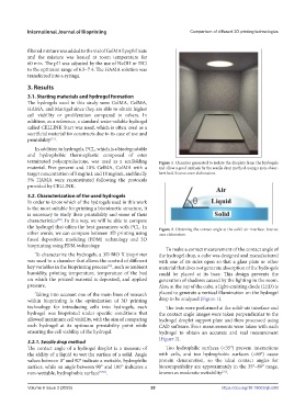
terminated polycaprolactone, was used as a scaffolding Figure 1. Chamber generated to isolate the droplets from the hydrogels
material. Five percent and 10% GelMA, ColMA with a and allow a good analysis by the sessile drop method, using a non-absor-
target concentration of 5 mg/mL and 10 mg/mL, and finally bent bed. Source: own elaboration.
5% HAMA were reconstituted following the protocols
provided by CELLINK.
3.2. Characterization of the used hydrogels
In order to know which of the hydrogels used in this work
is the most suitable for printing a biomimetic structure, it
is necessary to study their printability and some of their
[28]
characteristics . In this way, we will be able to compare
the hydrogel that offers the best guarantees with PCL. In Figure 2. Obtaining the contact angle at the solid–air interface. Source:
other words, we can compare between 3D printing using own elaboration.
fused deposition modeling (FDM) technology and 3D
bioprinting using FDM technology.
To make a correct measurement of the contact angle of
To characterize the hydrogels, a 3D BIO X bioprinter the hydrogel drop, a cube was designed and manufactured
was used in a chamber that allows the control of different with one of its sides open so that a glass plate or other
key variables in the bioprinting process , such as ambient material that does not generate absorption of the hydrogels
[29]
humidity, printing temperature, temperature of the bed could be placed at its base. This design prevents the
on which the printed material is deposited, and applied generation of shadows caused by the lighting in the room.
pressure. Also, at the top of the cube, a light-emitting diode (LED) is
Taking into account one of the main lines of research placed to generate a vertical illumination on the hydrogel
within bioprinting is the optimization of 3D printing drop to be analyzed (Figure 1).
technology for introducing cells into hydrogels, each The tests were performed at the solid–air interface and
hydrogel was bioprinted under specific conditions that the contact angle images were taken perpendicular to the
allowed maximum cell viability, with the aim of comparing hydrogel droplet support plate and then processed using
each hydrogel at its optimum printability point while CAD software. Four measurements were taken with each
ensuring the cell viability of the hydrogel. hydrogel to obtain an accurate and real measurement
(Figure 2).
3.2.1. Sessile drop method
The contact angle of a hydrogel droplet is a measure of Too hydrophilic surfaces (<35°) prevent interactions
the ability of a liquid to wet the surface of a solid. Angle with cells, and too hydrophobic surfaces (>80°) cause
values between 0° and 90° indicate a wettable, hydrophilic protein denaturation, so the ideal contact angles for
surface, while an angle between 90° and 180° indicates a biocompatibility are approximately in the 35°–80° range,
[31]
non-wettable, hydrophobic surface [9,30] . known as moderate wettability .
Volume 9 Issue 3 (2023) 29 https://doi.org/10.18063/ijb.680