
Page 311 - IJB-9-4
P. 311
International Journal of Bioprinting Biomechanical properties of 3D printable materialv
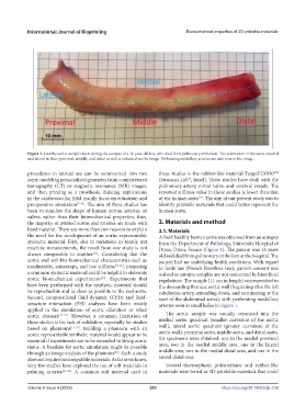
Figure 1. Healthy aortic sample taken during the autopsy of a 16-year-old boy who died from pulmonary embolism. The orientation of the aorta (medial
and lateral in blue; proximal, middle, and distal in red) is indicated on the image. Perforating medullary arteries are also seen in the image.
procedures in iatrical use can be summarized into two these studies is the rubber-like material TangoFLX930
TM
steps: modeling personalized geometry from computerized (Stratasys Ltd. , Israel). These studies have dealt with the
©
tomography (CT) or magnetic resonance (MR) images, pulmonary artery, mitral valve, and cerebral vessels. The
and then printing as a prosthesis. Existing applications reported stiffness value in these studies is lower than that
in the cardiovascular field mainly focus on education and of the human aorta . The aim of our present study was to
[11]
preoperative simulation [7-10] . The aim of these studies has identify printable materials that could better represent the
been to simulate the shape of human aortas, arteries, or human aorta.
valves, rather than their biomechanical properties; thus,
the majority of printed aortas and arteries are made with 2. Materials and method
hard material. There are more than two reasons to explain 2.1. Materials
the need for the development of an aortic representable A fresh healthy human aorta was obtained from an autopsy
syntactic material. First, due to variations in tensile test from the Department of Pathology, University Hospital of
machine measurements, the result from one study is not Dijon, Dijon, France (Figure 1). The patient was 16 years
always comparable to another . Considering that the old and died from pulmonary embolism at the hospital. The
[11]
aortic wall exhibits biomechanical characteristics such as patient had no underlying health conditions. With regard
nonlinearity, anisotropy, and low stiffness [11,12] , proposing to Jardé law (French Bioethics law), patient consent was
a common syntactic material could be helpful to elaborate waived as autopsy samples are not concerned by bioethical
aortic biomechanical experiments . Experiments that regulations. The sample (11 cm in length) corresponded to
[13]
have been performed with the synthetic material should the descending thoracic aortic wall (beginning after the left
be reproducible and as close as possible to the real aorta. subclavian artery, extending down, and terminating at the
Second, computational fluid dynamic (CFD) and fluid- start of the abdominal aorta), with perforating medullary
structure interaction (FSI) analyses have been widely arteries seen as small holes in Figure 1.
applied in the simulation of aortic dilatation or other
aortic diseases [14-16] . However, a common limitation of The aortic sample was visually separated into the
these studies is the lack of validation, especially for studies medial aortic quadrant (smaller curvature of the aortic
based on phantoms [17-19] . Building a phantom with an wall), lateral aortic quadrant (greater curvature of the
aortic representable synthetic material would appear to be aortic wall), proximal aorta, middle aorta, and distal aorta.
essential if experiments are to be extended to living aortic Six specimens were obtained: one in the medial proximal
tissue. A baseline for aortic simulation might be possible area, two in the medial middle area, one in the lateral
through an image analysis of the phantom . Such a study middle area, one in the medial distal area, and one in the
[20]
does not require biocompatible materials. As far as we know, lateral distal area.
very few studies have explored the use of soft materials in Several thermoplastic polyurethane and rubber-like
printing arteries [21-24] . A common soft material used in materials were tested as 3D-printable materials that could
Volume 9 Issue 4 (2023) 303 https://doi.org/10.18063/ijb.736