
Page 402 - IJB-9-4
P. 402
International Journal of Bioprinting Impingement shear stress during microvalve-based bioprinting
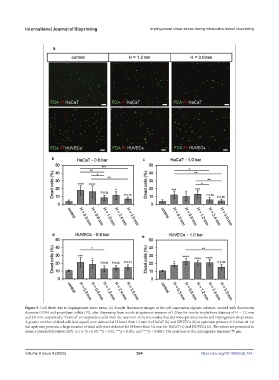
Figure 9. Cell death due to impingement shear stress. (a) Sample fluorescent images of the cell-suspension alginate solution, stained with fluorescein
diacetate (FDA) and propidium iodide (PI), after dispensing from nozzle at upstream pressure of 1.0 bar for nozzle-to-platform distance of H = 1.2 mm
and 3.0 mm, respectively. “Control” corresponds to cells from the reservoir of the microvalve that did not experience nozzle and impingement shear stress.
A greater number of dead cells (red signal) were detected at H lower than 1.2 mm for HaCaT (b) and HUVECs (d) at upstream pressure of 0.6 bar. At 1.0
bar upstream pressure, a large number of dead cells were detected for H lower than 3.0 mm for HaCaT (c) and HUVECs (e). The values are presented as
mean ± standard deviation (SD). n ≥ 3, *p < 0.05, **p < 0.01, ***p < 0.001, and ****p < 0.0001. The scale bars in the micrographs represent 75 µm.
Volume 9 Issue 4 (2023) 394 https://doi.org/10.18063/ijb.743