
Page 403 - IJB-9-4
P. 403
International Journal of Bioprinting Impingement shear stress during microvalve-based bioprinting
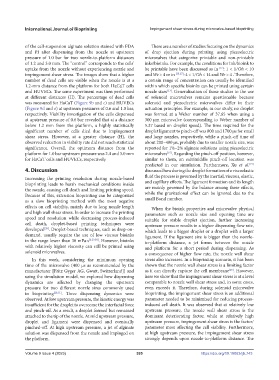
of the cell-suspension alginate solution stained with FDA There are a number of studies focusing on the dynamics
and PI after dispensing from the nozzle at upstream of drop ejection during printing using piezoelectric
pressure of 1.0 bar for two nozzle-to-platform distances microvalves that categorize printable and non-printable
of 1.2 and 3.0 mm. The “control” corresponds to the cells’ inks/bioinks. For example, the conditions for ink/bioink to
uptake from the nozzle without experiencing nozzle and be printable have been discussed as in [12] : 1 < 1 ⁄ Oh < 10
impingement shear stress. The images show that a higher and We > 4 or in [13,27] : 4 < 1 ⁄ Oh < 14 and We > 4 . Therefore,
number of dead cells are visible when the nozzle is at a a certain range of concentration can usually be identified
1.2-mm distance from the platform for both HaCaT cells within which specific bioinks can be printed using certain
and HUVECs. The same experiment was then performed nozzle sizes . Generalization of those studies to the use
[13]
at different distances (H). The percentage of dead cells of solenoid microvalves remains questionable because
was measured for HaCaT (Figure 9b and c) and HUVECs solenoid and piezoelectric microvalves differ in their
(Figure 9d and e) at upstream pressures of 0.6 and 1.0 bar, actuation principles. For example, in our study, no droplet
respectively. Viability investigation of the cells dispensed was formed at a Weber number of 37.65 when using a
at upstream pressure of 0.6 bar revealed that at a distance 300 µm microvalve (corresponding to Weber number of
below 1.2 mm from the platform, a highly statistically 5.37 based on droplet speed). The time required for the
significant number of cells died due to impingement droplet ligament to pinch-off was 800 and 1700 µs for small
shear stress. However, at a greater distance (H), the and large nozzles, respectively, while a pinch-off time of
observed reduction in viability rate did not reach statistical about 200–400 µs, probably due to smaller nozzle size, was
significance. Overall, the optimum distance from the reported for 1%–2% alginate solutions using piezoelectric
platform for 1.0 bar upstream pressure was 2.4 and 3.0 mm microvalves . Regarding the pinch-off position, however,
[15]
for HaCaT cells and HUVECs, respectively. similar to them, an exit/middle pinch-off location was
predicted in our simulation. Furthermore, Xu et al.
[15]
4. Discussion discussed how during the droplet formation of a viscoelastic
Increasing the printing resolution during nozzle-based fluid the process is governed by the inertial, viscous, elastic,
bioprinting leads to harsh mechanical conditions inside and capillary effects. The ligament formation and its length
the nozzle, causing cell death and limiting printing speed. are mainly governed by the balance among these effects,
Because of this, extrusion bioprinting can be categorized while the gravitational effect can be ignored due to the
as a slow bioprinting method with the most negative small Bond number.
effects on cell viability, mainly due to long nozzle length When the bioink properties and microvalve physical
and high wall shear stress. In order to increase the printing parameters such as nozzle size and opening time are
speed and resolution while decreasing process-induced suitable for stable droplet ejection, further increasing
cell death, droplet-based printing techniques were upstream pressure results in a higher dispensing flow rate,
developed . Droplet-based techniques, such as drop-on- which leads to a bigger droplet or a droplet with a larger
[28]
demand, usually require the use of low viscous bioinks ligament. If the ligament size is bigger than the nozzle-
in the range lower than 30 mPa·s [2,29,30] . However, bioinks to-platform distance, a jet forms between the nozzle
with relatively higher viscosity can still be printed using and platform for a short period during dispensing. As
solenoid microvalves. a consequence of higher flow rate, the nozzle wall shear
In this work, considering the minimum opening stress also increases. In a bioprinting scenario, it has been
time of the microvalve (400 µs as recommended by the shown that the nozzle wall shear stress is a limiting factor
manufacturer [Fritz Gyger AG, Gwatt, Switzerland]) and as it can directly rupture the cell membrane . However,
[25]
using the simulation model, we explored how dispensing here we show that the impingement shear stress is at a level
dynamics are affected by changing the upstream comparable to nozzle wall shear stress and, in some cases,
pressure for two different nozzle sizes commonly used even exceeds it. Therefore, during solenoid microvalve
in bioprinting [20,31] . Three dispensing dynamics were bioprinting, the impingement shear stress is an additional
observed. At low upstream pressure, the kinetic energy was parameter needed to be minimized for reducing process-
insufficient for the droplet to overcome the interfacial force induced cell death. It was observed that at relatively low
and pinch-off. As a result, a droplet formed but remained upstream pressure, the nozzle wall shear stress is the
attached to the tip of the nozzle. At mid upstream pressure, dominant deteriorating factor, while at relatively high
droplet and ligament were dispensed and eventually upstream pressure, impingement shear stress is the factor/
pinched-off. At high upstream pressure, a jet of alginate parameter most affecting the cell viability. Furthermore,
solution was dispensed from the nozzle and impinged on at high upstream pressure, the impingement shear stress
the platform. strongly depends upon nozzle-to-platform distance. The
Volume 9 Issue 4 (2023) 395 https://doi.org/10.18063/ijb.743