
Page 122 - IJB-9-5
P. 122
International Journal of Bioprinting DLP-printed scaffold for bone regeneration
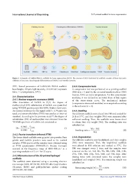
Figure 1. Schematic of GelMA/PMAA scaffolds for bone regeneration (ECO). The process of ECO facilitated by scaffolds consists of three key parts:
chelation of iron ions, chondrogenic differentiation of BMSCs, and vascular invasion.
29°C. Kernel parameters of GelMA/6% PMAA scaffold: 2.4.4. Compressive tests
layer height, 100 μm; light intensity, 10 mW/cm ; exposure A compression test was performed on a printing scaffold
2
time, 11 s; temperature, 29°C). (Ф10 mm × 5 mm) by the universal tensile machine (3365
Instron, USA) at room temperature. For the compression
2.4. Characterization modulus, it was defined as an initial slope of line region
2.4.1. Nuclear magnetic resonance (NMR) of the stress–strain curve. The mechanical indexes
After dissolution of GelMA in D O, the degree of (compressive stress and modulus) were acquired according
2
methacryloyl (DS) substitution of GelMA was quantified to the software (n = 3).
using H NMR spectroscopy (400 MHz, Bruker, Germany).
1
An internal reference for the signal with δ = 4.79 ppm was 2.4.5. Swelling
used, and tetramethylsilane (TMS) was used as an internal The different scaffolds were placed into PBS and soaked for
standard. According to the previous study , the degree of 24 h at 37°C, and their weights (Ws) were measured after
[44]
substitution (DS) of methacrylate was obtained from the sufficient swelling. Then, the scaffolds were freeze-dried
1 H NMR spectrum of GelMA and calculated as: to obtain their dry weight (Wd). The swelling ratio was
calculated as:
.
0 3462 I
DS 55. ppm (I) Ws Wd
0 0380. I Swelling ratio (II)
1ppm Wd
2.4.2. Fourier transform infrared (FTIR)
The freeze-dried scaffolds were grinded into powder. Pure 2.4.6. Degradation
gelatin and GelMA powders were used as the control The different scaffolds were lyophilized, and their weights
samples. FTIR spectra of the samples were obtained using (W0) were measured. Then, the lyophilized scaffolds
an FTIR spectrometer (TENSOR-27, Bruker, German) were placed in PBS solution and soaked at 37°C. The
scanned in the frequency range of 4000–500 cm at a PBS was changed every 2 days, and the samples were
-1
scanning resolution of 2 cm for 32 scans. removed on the 1st, 3rd, 5th, 7th, 9th, 12th, 15th, 21th,
-1
27th, 33th, 39th, 45th, 51th, 54th, and 55th days. After
2.4.3 Microstructure of the 3D-printed hydrogel rinsing twice with deionized water, the samples were
scaffolds lyophilized and weighed (Wt). The remaining weight was
The scaffolds were observed using a scanning electron calculated as:
microscope (SEM, HITACHI, SU8100) after lyophilization
(Quorum, K850) and gold/palladium sputter coating Remaining weight % Wt 100% (III)
(HITACHI, MC1000). W0
Volume 9 Issue 5 (2023) 114 https://doi.org/10.18063/ijb.754