
Page 127 - IJB-9-5
P. 127
International Journal of Bioprinting DLP-printed scaffold for bone regeneration
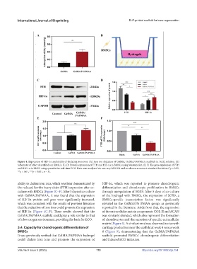
Figure 4. Expression of HIF-1α and ability of chelating iron ions. (A) Iron ion chelation of GelMA, GelMA/3%PMAA scaffolds in FeCl solution. (B)
3
Schematic of effect of scaffolds on BMSCs. (C, D) Protein expression of FTH and HIF-1α in BMSCs using Western blot. (E, F) The gene expression of FTH
and HIF-1α in BMSC using quantitative real-time PCR. Data were analyzed via one-way ANOVA and are shown as mean ± standard deviation (*p < 0.05,
**p < 0.01, ***p < 0.001, n = 3).
ability to chelate iron ions, which was later demonstrated by HIF-1α, which was reported to promote chondrogenic
the reduced ferritin heavy chain (FTH) expression after co- differentiation and chondrocyte proliferation in BMSCs
culture with BMSCs (Figure 4C–F). After 5 days of co-culture through upregulation of SOX9. After 5 days of co-culture
with GelMA/3%PMAA, it was found that the expression of the hydrogel with BMSCs, the expression of SOX9, a
of HIF-1α protein and gene were significantly increased, BMSCs-specific transcription factor, was significantly
which was consistent with the results of previous literature elevated in the GelMA/3% PMAA group, as previously
that the reduction of iron ions could promote the expression reported in the literature. Aside from that, the expression
of HIF-1α (Figure 4C–F). These results showed that the of the extracellular matrix components COL-II and ACAN
GelMA/3%PMAA scaffold could play a role similar to that was similarly elevated, which also represent the formation
of a low oxygen environment, providing the basis for ECO. of chondrocytes and the secretion of specific extracellular
matrix (Figure 5). A similar trend was observed in vivo with
3.4. Capacity for chondrogenic differentiation of cartilage production near the scaffold at week 4 versus week
BMSCs 8 (Figure 9), demonstrating that the GelMA/3%PMAA
It was previously verified that GelMA/3%PMAA hydrogel scaffold promoted BMSCs’ chondrogenic differentiation
could chelate iron ions and promote the expression of and induced ECO initiation.
Volume 9 Issue 5 (2023) 119 https://doi.org/10.18063/ijb.754