
Page 129 - IJB-9-5
P. 129
International Journal of Bioprinting DLP-printed scaffold for bone regeneration
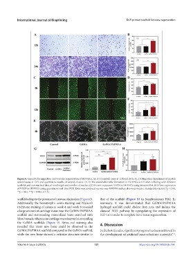
Figure 6. Capacity for migration and vascular regeneration of HUVECs. (A, B) Transwell assay at 12 h and 24 h. (C, D) Migration experiment of scratch
wound assay at 12 h and quantitative results of wound closure. (E, F) The endothelial tube formation in HUVECs at 6 h after culturing with different
scaffolds and summarized data of total length and number of meshes. (G) Protein expression VEGF in HUVECs using Western blot. (H) Gene expression
of VEGF in HUVECs using quantitative real-time PCR. Data were analyzed via one-way ANOVA and are shown as mean ± standard deviation (*p < 0.05,
**p < 0.01, ***p < 0.001, n = 3).
scaffolds despite the presence of neovascularization (Figure 8). that of the scaffold (Figure S3 in Supplementary File). In
Additionally, the hematoxylin–eosin staining and Masson’s summary, it was demonstrated that GelMA/3%PMAA
trichrome staining of tissues at week 4 and week 8 revealed hydrogel scaffold could chelate iron ions and induce the
a large amount of cartilage tissue near the GelMA/3%PMAA classical ECO pathway by upregulating the expression of
scaffold and surrounding mineralized bone enriched with HIF-1α in order to complete bone tissue regeneration.
blood vessels, whereas no cartilage was observed surrounding
the GelMA scaffolds (Figure 9). Sirius red staining also 4. Discussion
revealed that more new bone could be observed in the
GelMA/3%PMAA scaffold compared to the GelMA scaffold, In the last decades, significant progress has been achieved in
while the new bone showed a reticular structure similar to the development of artificial bone-substitute materials .
[45]
Volume 9 Issue 5 (2023) 121 https://doi.org/10.18063/ijb.754