
Page 126 - IJB-9-5
P. 126
International Journal of Bioprinting DLP-printed scaffold for bone regeneration
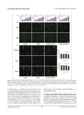
Figure 3. Biocompatibility of GelMA/PMAA scaffold in vitro. (A) CCK-8 assay showing the proliferation of BMSCs after co-cultured with different
extracts of different scaffolds for 1–5 days. (B) Live/dead assay of BMSCs co-cultured with 25% extract; green represents living cells and red represents
dead cells. (C) Phalloidin assay of BMSCs co-cultured with 25% extract. (D) Number of cells in live/dead assay. Data were analyzed via one-way ANOVA
and are shown as mean ± standard deviation (*p < 0.05, **p < 0.01, ***p < 0.001, n = 3).
of PMAA (Figure 3A). Therefore, the extract with 25% of Supplementary File). Therefore, GelMA/3%PMAA is a
concentration was selected for the next immunofluorescence more suitable bio-ink.
staining. Live/dead staining showed a poor proliferation of
cells cultured in the GelMA/6% PMAA extract (Figure 3B 3.3. Expression of HIF-1α after chelation of iron ions
and D). In contrast, the morphology and proliferation of In hypoxic environments, a series of reactive behaviors are
the cells were not significantly affected by the extract of performed by cells, such as elevation of HIF-1α, a signal
GelMA/3%PMAA (Figure 3C). The cells on either GelMA molecule that senses oxygen, which subsequently triggers a
or GelMA/3% PMAA scaffold showed high viability after series of biological effects. As shown in Figure 4A, it could
24 h of inoculation on the scaffold surface (Figure S2 in be found that GelMA/3%PMAA hydrogel possessed the
Volume 9 Issue 5 (2023) 118 https://doi.org/10.18063/ijb.754