
Page 125 - IJB-9-5
P. 125
International Journal of Bioprinting DLP-printed scaffold for bone regeneration
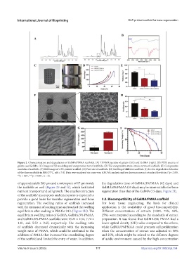
Figure 2. Characterization and degradation of GelMA/PMAA scaffolds. (A) H NMR spectra of gelatin (left) and GelMA (right). (B) FTIR spectra of
1
gelatin and GelMA. (C) Images of 3D modeling and compression test of scaffolds. (D) The compressive stress–strain curves of scaffolds. (E) Compressive
modulus of scaffolds. (F) SEM image of a 3D-printed scaffold. (G) Pore size of scaffolds. (H) Swelling of different scaffolds. (I) In vitro degradation behavior
of the three scaffolds in PBS (37°C, pH = 7.4). Data were analyzed via a one-way ANOVA analysis and are shown as mean ± standard deviation (*p < 0.05,
**p < 0.01, ***p < 0.001, n = 3).
of approximately 500 μm and a micropore of 17 μm inside the degradation time of GelMA/3%PMAA (42 days) and
the scaffolds as well (Figure 2F and G), which facilitated GelMA/6% PMAA (55 days) may be more suitable for bone
nutrient transport and cell growth. The excellent structure regeneration than that of the GelMA (15 days, Figure 2I).
of the scaffolds’ macropores and micropores is expected to
provide a good basis for vascular regeneration and bone 3.2. Biocompatibility of GelMA/PMAA scaffold
regeneration. The swelling ratios of scaffolds increased For bone tissue engineering, the basis for clinical
with the extension of soaking time and reached the swelling application is the availability of good biocompatibility.
equilibrium after soaking in PBS for 24 h (Figure 2H). The Different concentrations of extracts (100%, 75%, 50%,
equilibrium swelling ratios of GelMA, GelMA/3% PMAA, 25%) were prepared according to the standards of extract
and GelMA/6% PMAA scaffolds were 13.21 ± 2.52, 7.74 ± preparation. It was found that GelMA/6% PMAA had a
1.41, and 5.52 ± 0.65, respectively. The swelling ratio lower optical density (OD) value compared to the others,
of scaffolds decreased dramatically with the increasing while GelMA/3%PMAA could promote cell proliferation
weight ratio of PMAA, which could be attributed to the when the concentration of extract was adjusted to 50%
addition of PMAA that increased the crosslinking degree and 25%, which might be related to the different degrees
of the scaffold and limited the entry of water. In addition, of acidic environment caused by the high concentration
Volume 9 Issue 5 (2023) 117 https://doi.org/10.18063/ijb.754