
Page 128 - IJB-9-5
P. 128
International Journal of Bioprinting DLP-printed scaffold for bone regeneration
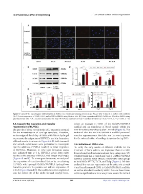
Figure 5. Capacity for chondrogenic differentiation of BMSCs. (A) Fluorescent staining of Col-II and ACAN after 5 days of co-culture with scaffolds.
(B, C) Protein expression of SOX9, Col-II, and ACAN in BMSCs using Western blot. (D) Gene expression of SOX9, Col-II, and ACAN in BMSCs using
quantitative real-time PCR. Data were analyzed via one-way ANOVA and are shown as mean ± standard deviation (*p < 0.05, **p < 0.01, ***p < 0.001, n = 3).
3.5. Capacity for migration and vascular where an increase in CD31 of the GelMA/3%PMAA
regeneration of HUVECs scaffold and an abundance of blood vessels within the
The growth of blood vessels in the ECO process is essential new bone tissue were found after 1 month (Figure 8). This
for the mineralization of cartilage templates. Therefore, indicated that the GelMA/3%PMAA scaffold promoted
we investigated the ability of GelMA/3%PMAA hydrogel vascular regeneration at the defect site that was important
to promote the migration of HUVECs and the formation for the mineralization of cartilage templates during ECO.
of blood vessels. As shown in Figure 6A–D, both transwell
and scratch experiments were performed to investigate 3.6. Initiation of ECO in vivo
that the addition of PMAA resulted in better migration To verify the early results of different scaffolds for the
of HUVECs. Moreover, in vitro tube formation assays treatment of bone defects, we implanted them in rabbit
also indicated that at 6 h, HUVECs could form more femoral condylar defects and analyzed them using micro-CT,
meshes in the presence of PMAA, with longer total length which revealed that at week 4 and week 8, GelMA/3%PMAA
(Figure 6E and F). To investigate the reason, we analyzed scaffolds achieved better efficacy compared to other groups
the expression of vascular-related factors by co-culturing in both BMD, BV/TV, Th.Tb, and Th.Sp (Figure 7). We then
HUVECs with hydrogel. GelMA/3%PMAA hydrogel was analyzed the vascular regeneration at the defect site at week
found to promote the protein and mRNA expression of 4 and found increased CD31 in GelMA/3%PMAA scaffolds
VEGF (Figure 6G and H). The scaffold was then implanted as well as abundant vascularity within the new bone tissue,
into the defect site of the rabbit femoral condyle bone, while no significant new bone was present around the GelMA
Volume 9 Issue 5 (2023) 120 https://doi.org/10.18063/ijb.754