
Page 176 - IJB-9-5
P. 176
International Journal of Bioprinting Functional materials of 3D bioprinting for wound healing
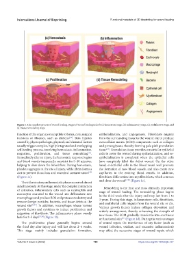
Figure 1. The complete process of wound healing. Stages of wound healing include (a) hemostasis stage, (b) inflammatory stage, (c) proliferative stage, and
(d) tissue remodeling stage.
function of this organ are susceptible to burns, cuts, surgical epithelialization, and angiogenesis. Fibroblasts migrate
incisions or illnesses, such as diabetes . Skin injuries from the surrounding tissue to the wound site to produce
[33]
caused by physiopathologic, physical, and chemical factors extracellular matrix (ECM) components such as collagen
usually trigger complex, highly integrated and overlapping and proteoglycans, thereby forming pale pink granulation
self-healing process, involving hemostasis, inflammation, tissue . Granulation tissue provides a matrix for epithelial
[39]
migration, proliferation, and tissue remodeling . cells to cover the wound during epithelialization, and re-
[34]
Immediately after an injury, the hemostatic response begins epithelialization is completed when the epithelial cells
and blood vessels temporarily constrict for 5–10 minutes, have completely filled the defect wound. On the other
helping to slow down the blood flow. During hemostasis, hand, endothelial cells in the blood vessel wall promote
platelets aggregate at the site of injury, while fibrin forms a the formation of new blood vessels, and also create new
clot to prevent blood loss and microbial contamination capillaries in the existing blood vessels. In addition,
[35]
(Figure 1a). fibroblasts differentiate into myofibroblasts, which contract
and close the wound [2,40] (Figure 1c).
The inflammatory and hemostatic phases occurred almost
simultaneously. At this stage, under the complex interaction Remodeling is the final and most clinically important
of cytokines, inflammatory cells such as neutrophils and stage of wound healing. The remodeling phase begins
monocytes recruited to the wound site differentiate into in the third week after the injury and may last from 1 to
macrophages and produce ROS and proteases to destroy and 3 years. During this stage, inflammatory cells, fibroblasts,
remove foreign particles, bacteria, and tissue debris at the and endothelial cells migrate from the wound site or die.
wound site [36,37] . In addition, macrophages release various Various growth factors induce collagen deposition and
growth factors and cytokines to induce proliferation and orderly arrangement, thereby enhancing the strength of
migration of fibroblasts. The inflammatory phase usually new tissue. The ECM gradually transforms into scar tissue
lasts for 2–5 days [35,38] (Figure 1b). or functional skin (Figure 1d). During the various stages
[41]
The proliferative phase generally begins around of wound repair, the interference of any factor (such as
the third day after injury and will last about 2–4 weeks. wound infection, oxidant, and excessive inflammation)
This stage mainly includes granulation formation, may affect the successive stages of wound repair, which
Volume 9 Issue 5 (2023) 168 https://doi.org/10.18063/ijb.757