
Page 316 - IJB-9-5
P. 316
International Journal of Bioprinting Scaffold for engineering enthesis organ
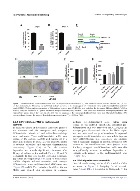
Figure 11. Proliferation and differentiation of MSCs on electrospun PLGA scaffolds (ePLGA). MSCs were seeded on different scaffolds for 3 (A) or 7
(B) days. In the end, the MTS assay was performed. Data are expressed as the percentage of cell proliferation versus undifferentiated MSCs seeded on
plastic (CTRL). (C) Schematic representation of differentiation protocol used. (D–F) Cells were seeded on the electrospun PLGA scaffolds (ePLGA) or
plastic (CTRL) and maintained in a growth medium or tenogenic medium (Teno) for 14 or 21 days. In the end, aniline blue staining was performed, and
representative images after 21 days were reported (D). Data are expressed as the percentage of differentiation versus non-differentiated (CTRL) MSCs
grown on plastic. Data are the results of three independent experiments. ***p < 0.001 vs. CTRL.
3.5.3. Differentiation of MSC on multimaterial medium (pre-differentiated MSC) before being
scaffolds seeded on the scaffold. Specifically, osteoblast pre-
To assess the ability of the enthesis scaffold to promote differentiated cells were seeded on the PCL region and
and maintain both the osteogenic and tenogenic tenocyte pre-differentiated cells on the PLGA region
differentiation, alizarin red and aniline blue stainings and then maintained in a growth medium. As expected,
were performed. Thus, undifferentiated MSCs were osteogenic pre-differentiated cells were able to improve
seeded on the enthesis scaffold and maintained in a their differentiation into osteoblasts on enthesis,
growth medium for 14 and 21 days to assess the ability with a significant increase of calcium deposition with
to support osteoblast and tenocyte differentiation, respect to the undifferentiated ones (Figure 12B).
respectively (Figure 12A). In fact, the calcium Similarly, tenogenic pre-differentiated cells were able
deposition was already significantly increased after to significantly increase the collagen deposition on
14 days of culture on the scaffold (Figure 10E and F). enthesis with respect to the undifferentiated ones
Conversely, 21 days were needed to obtain a significant (Figure 12C).
deposition of collagen (Figure 11E and F). The enthesis
scaffold slightly induced osteoblast and tenocyte 3.6. Clinically-relevant scale scaffold
differentiation when undifferentiated MSCs were used Uniaxial tensile testing results of 3D braided scaffolds
(Figure 12B and C). Then to induce the differentiation, are shown in Figure 13. Analyzing the stress–strain
MSCs were primed with osteogenic or tenogenic curve (Figure 13B), it is possible to distinguish: (i) the
Volume 9 Issue 5 (2023) 308 https://doi.org/10.18063/ijb.763