
Page 507 - IJB-9-5
P. 507
International Journal of Bioprinting Implantation of composites for cartilage repair
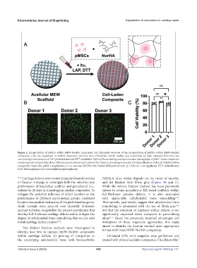
Figure 2. Encapsulation of pMSCs within MEW-NorHA composites. (A) Schematic overview of the encapsulation of pMSCs within MEW-NorHA
composites. Cells are suspended in NorHA macromer, which is then infilled into MEW meshes and crosslinked via light-mediated (hν) thiol-ene
crosslinking in the presence of LAP photoinitiator and DTT crosslinker. (B) Live/Dead staining and representative micrographs of pMSC-laden composites
containing cells sourced from three different porcine donors and cultured for 7 days in chondrogenic media. (C) Quantification of the cell viability within
composites 7 days after pMSC encapsulation. n = 6, one-way ANOVA with Tukey’s HSD post-hoc test, *p < 0.05, ns = not significant. DTT: dithiothreitol;
LAP: lithium phenyl-2,4,6-trimethylbenzoylphosphinate.
[10] . Cartilage defects were created along the femoral trochlea PLDLLA pins, which degrade on the order of months,
of Yucatan minipigs to investigate both the retention and and (ii) fixation with fibrin glue (Figure 5B and C).
performance of implanted acellular and precultured (i.e., While the former fixation method has been previously
culture for 28 days in chondrogenic media) composites. To shown to retain nanofibrous HA-based scaffolds within
mitigate the potential influence of defect location on the full-thickness porcine defects, it is also associated
[16]
performance of different experimental groups, treatment with appreciable subchondral bone remodeling .
location was randomized across all the performed surgeries. Alternatively, past results suggest that subchondral bone
[16]
Adult animals were selected over skeletally immature remodeling is attenuated with the use of fibrin glue
animals to better recapitulate the patient populations that but that the retention of implants within defects is not
develop full-thickness cartilage defects and to mitigate the significantly improved when compared to press-fitting
degree of subchondral bone remodeling that occurs with alone . Given the previously reported advantages and
[17]
[39]
initial cartilage defect creation . limitations of these respective approaches, this study
aimed to identify the fixation method most appropriate
Two distinct fixation methods were investigated to
identify how best to implant MEW-NorHA composites for use with these MEW-NorHA composites.
within cartilage defects: (i) pinning of composites to Unilateral stifle joint surgeries were performed and
the underlying subchondral bone with bioresorbable treated with pinned acellular composites (“Acellular+Pin,”
Volume 9 Issue 5 (2023) 499 https://doi.org/10.18063/ijb.775