
Page 511 - IJB-9-5
P. 511
International Journal of Bioprinting Implantation of composites for cartilage repair
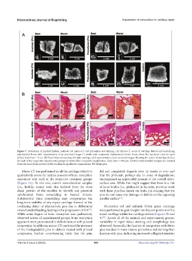
Figure 7. Evaluation of implant fixation methods via micro-CT and picrosirius red staining. (A) Micro-CT scans of cartilage defects and underlying
subchondral bone, with representative cross-sectional images 12 weeks after composite implantation shown. Insets show the top-down view for each
defect. Scale bars = 1 cm. (B) Picrosirius red staining of repair cartilage, with representative cross-sectional images showing the center of cartilage defects
for each of the respective experimental groups 12 weeks after composite implantation. Scale bars = 500 mm. Control osteochondral samples are isolated
from the most distal portion of the trochlea for qualitative comparisons. FG: fibrin glue.
Micro-CT was performed on all the cartilage defects to did not completely degrade over 12 weeks in vivo and
qualitatively assess the relative amount of bone resorption that the pinheads, perhaps due to onset of degradation,
associated with each of the respective treatment groups encompassed an appreciable amount of the overall defect
(Figure 7A). To this end, control osteochondral samples surface area. While this might suggest that there is a risk
(i.e., healthy tissue) were also isolated from the most of loose bodies (i.e., pinheads) in the joint, previous work
distal portion of the trochlea to identify any potential with these pins has shown via India ink staining that the
subchondral bone remodeling in treated defects. pins do not cause any damage or defects on the opposing
Subchondral bone remodeling may compromise the patellar surface .
[16]
long-term stability of any repair cartilage formed in the
overlaying defect or alternatively give rise to differential Picrosirius red and safranin O/fast green stainings
osteochondral loading, leading to the progression of OA . were performed to gain insight into the composition of the
[42]
While some degree of bone resorption was qualitatively repair cartilage within the cartilage defects (Figures 7B and
observed across all experimental groups, bone resorption 8) . Across all of the animals and experimental groups,
[32]
appeared more pronounced in defects treated with pinned variability in repair tissue staining and morphology was
composites. In addition, micro-CT confirmed the presence observed. Generally, the fixation of composites with fibrin
of the biodegradable pins in defects treated with pinned glue resulted in more intense picrosirius red staining than
composites, further corroborating both that the pins fixation with pins, indicating increased collagen formation
Volume 9 Issue 5 (2023) 503 https://doi.org/10.18063/ijb.775