
Page 509 - IJB-9-5
P. 509
International Journal of Bioprinting Implantation of composites for cartilage repair
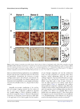
Figure 4. ECM staining of composites across pMSC donors. After 28 days of culture in chondrogenic media, ECM within composites containing
encapsulated pMSCs from three porcine donors is visualized via (A) alcian blue staining (for sulfated glycosaminoglycans, sGAG), (B) type II collagen
(COL II) immunohistochemistry, and (C) type I collagen (COL I) immunohistochemistry. (A–C) Representative images (left) and quantification of
staining intensity (right) to characterize (A) sGAG, (B) COL II, and (C) COL I distribution and organization for three porcine donors. n = 81 images, 9
sections, 3 composites; one-way ANOVA with Tukey’s HSD post-hoc test, **p < 0.01, ****p < 0.0001, ns = not significant.
failure was observed during implantation, it is possible that of pins through composites and into the subchondral
the pin head may have translocated away from the defect bone) may have perturbed the composite–native tissue
post-operatively. Alternatively, the formation of repair interface, further influencing defect fill and repair
cartilage around and/or above the pin could obscure the cartilage integration. Improved defect fill and macroscopic
presence of underlying pins. For example, pinheads were appearance were observed for composites fixed within
observed in both lateral defects in one animal much further defects using fibrin glue in lieu of pins; however, at
away from the articular surface (i.e., deeper) than in other least one defect appeared to be entirely empty (lateral
samples. proximal defect) (Figure 6). Specifically, both acellular
and precultured composites implanted using fibrin glue
Generally, macroscopic visualization of the trochlea exhibited instances where complete defect fill was achieved,
and arthroscopic imaging revealed that the volume of and a homogenous, smooth cartilage surface was observed;
the pin used to fix composites qualitatively impeded in parallel, other defects were partially filled at lesser
[30]
the complete filling of defects containing acellular or depths with more apparent fissures . Prior observations
precultured composites (Figure 6). Moreover, the potential for implants in ponies showed that fibrin was insufficient
contraction of composites upon formation of pilot holes to fix chondral implants, which indicates that the species,
[12]
or the application of the fixation guide (for the insertion as well as the implant location, must be considered .
Volume 9 Issue 5 (2023) 501 https://doi.org/10.18063/ijb.775