
Page 513 - IJB-9-5
P. 513
International Journal of Bioprinting Implantation of composites for cartilage repair
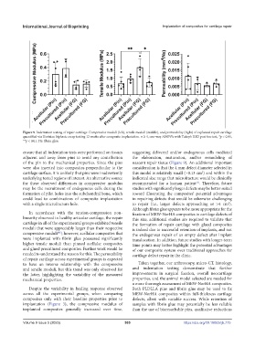
Figure 9. Indentation testing of repair cartilage. Compressive moduli (left), tensile moduli (middle), and permeability (right) of explanted repair cartilage
quantified via Hertzian biphasic creep testing 12 weeks after composite implantation. n ³ 4, one-way ANOVA with Tukey’s HSD post-hoc test, *p < 0.05,
**p < 0.01. FG: fibrin glue.
ensure that all indentation tests were performed on tissues suggesting delivered and/or endogenous cells mediated
adjacent and away from pins to avoid any contribution the elaboration, maturation, and/or remodeling of
of the pin to the mechanical properties. Since the pins nascent repair tissue (Figure 9). An additional important
were also inserted into composites perpendicular to the consideration is that the 4 mm defect diameter selected in
cartilage surface, it is unlikely that pins were inadvertently this model is relatively small (~0.13 cm ) and within the
2
underlying tested regions of interest. An alternative source indicated size range that microfracture would be clinically
for these observed differences in compressive modulus recommended for a human patient . Therefore, future
[1]
may be the recruitment of endogenous cells during the studies with significantly larger defects may be better suited
formation of pilot holes into the subchondral bone, which toward illustrating the composites’ potential advantages
could lead to combinations of composite implantation in repairing defects that would be otherwise challenging
with a single microfracture hole. to repair (i.e., larger defects approaching or >4 cm ).
2
Although fibrin glue appears to be more appropriate for the
In accordance with the tension-compression non- fixation of MEW-NorHA composites in cartilage defects of
linearity observed in healthy articular cartilage, the repair this size, additional studies are required to validate that
cartilage in all of the experimental groups exhibited tensile the formation of repair cartilage with glued composites
moduli that were appreciably larger than their respective is indeed due to successful retention of implants, and not
compressive moduli ; however, acellular composites that the endogenous repair of an empty defect after implant
[45]
were implanted with fibrin glue possessed significantly translocation. In addition, future studies with longer-term
higher tensile moduli than pinned acellular composites time points may better highlight the potential advantages
and glued precultured composites. Further work would be of our composite system over traditional approaches for
needed to understand the reason for this. The permeability cartilage defect repair in the clinic.
of repair cartilage across experimental groups is expected
to have an inverse relationship with the compressive Taken together, our arthroscopy, micro-CT, histology,
and tensile moduli, but this trend was only observed for and indentation testing demonstrate that further
the latter, highlighting the variability of the measured improvements in surgical fixation, overall neocartilage
mechanical properties. properties, and the animal model selected are needed for
a more thorough assessment of MEW-NorHA composites.
Despite the variability in healing response observed Both PLDLLA pins and fibrin glue may be used to fix
across all the experimental groups, when comparing MEW-NorHA composites within full-thickness cartilage
composites only with their baseline properties prior to defects, albeit with variable success. While retention of
implantation (Figure 3), the compressive modulus of samples with fibrin glue may potentially be less reliable
implanted composites generally increased over time, than the use of bioresorbable pins, qualitative reductions
Volume 9 Issue 5 (2023) 505 https://doi.org/10.18063/ijb.775