
Page 512 - IJB-9-5
P. 512
International Journal of Bioprinting Implantation of composites for cartilage repair
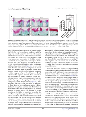
Figure 8. Evaluation of implant fixation methods via safranin O/fast green staining. (A) Safranin O/fast green staining of repair cartilage, with representative
cross-sectional images showing the center of cartilage defects for each of the respective experimental groups 12 weeks after composite implantation.
Control osteochondral samples were isolated from the most distal portion of the trochlea for qualitative comparisons. Scale bars = 500 mm. (B) ICRS II
scoring of repair cartilage by 3 blinded reviewers on a scale of 0–100 (worst–best), where each symbol (circle, square, and triangle) corresponds to scores
from each reviewer. n ³ 12, one-way ANOVA with Tukey’s HSD post-hoc test, **p < 0.01, ***p < 0.001, ****p < 0.0001. FG: fibrin glue.
within defects. In addition, picrosirius red staining revealed animal model, cell-free implants showed abundant cell
that fibrin glue-fixed composites exhibited improved defect ingrowth and similar results as cell-containing implants .
[43]
filling over pinned composites. However, only marginal Importantly, the degradation of MEW-NorHA composites
differences in picrosirius red staining intensity and tissue in vivo via hyaluronidase activity may similarly facilitate
morphology were observed when comparing acellular cell ingrowth, even in acellular composites, such that over
versus precultured composites. In healthy explanted time the scaffold is remodeled into de novo cartilage .
[37]
osteochondral tissues, safranin O and fast green staining Taken together with our results, we postulated that
was dark and robust, suggesting the abundant presence mechanical stability is more determining for the success of
of both proteoglycans and collagen within the cartilage the implant than the presence of cells and/or precultured
and bone phases of the tissue (Figure 8A). In some of the extracellular matrix.
fibrin glue-fixed composites, the integration of nascent Finally, the functional properties of repair cartilage
tissue was visualized with the adjacent healthy tissue. formed in treated defects were evaluated via indentation
However, pinned acellular and precultured composites creep testing, which enables the in situ mechanical testing
exhibited marginal safranin O staining, even directly of tissues within defects to elucidate the compressive
adjacent to the apparent pinhead. These observations modulus, tensile modulus, and permeability (Figure 9). The
were corroborated by ICRS II histological scoring, which average compressive modulus of repair cartilage across all
indicated that the fixation of composites with fibrin glue the experimental groups did not exceed 0.4 MPa, suggesting
significantly improved the quality of resultant repair that the repair cartilage possessed inferior mechanical
cartilage compared to the pin fixation (Figure 8B). When properties when compared to previously reported modulus
composites were fixed within defects via the application values for native cartilage . Indentation testing of healthy
[44]
of fibrin glue, the best-performing samples exhibited tissue controls isolated from the most distal region of the
significant proteoglycan content and distribution, as lateral trochlear groove (compressive modulus = 2.00 ±
evidenced by safranin O staining approaching what was 0.64 MPa) confirmed that each treatment group led to only
observed for control tissues. The safranin O staining partial restoration of the defect’s biomechanical function.
intensity was also higher in precultured composites than
in acellular composites, suggesting that the formation of While no statistical differences in compressive
nascent tissue prior to implantation via the chondrogenic modulus were observed between each of the treatment
preculture period may help to mediate repair cartilage groups, pinned composites, on average, possessed higher
maturation. However, tissue morphology consistent compressive moduli than glued composites. However, the
with fibrocartilage was observed across all defects [27,32] . elevated compressive moduli observed for pinned acellular
In another recent study that employed MEW-reinforced and precultured composites may be due to the presence of
hydrogels as osteochondral implants in an orthotopic large the pin within the defect. Careful attention was given to
Volume 9 Issue 5 (2023) 504 https://doi.org/10.18063/ijb.775