
Page 510 - IJB-9-5
P. 510
International Journal of Bioprinting Implantation of composites for cartilage repair
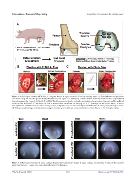
Figure 5. Study design to evaluate MEW-NorHA composite fixation in a porcine model of articular cartilage repair. (A) Full-thickness cartilage defects
are created along the trochlear groove of the patellofemoral joint (right, hind stifle joint). Defects are then filled with either acellular or precultured
(chondrogenic media, Donor 3 pMSCs, 28 days) MEW-NorHA composites. Twelve weeks after implantation, the retention of implants and the quality of
repair cartilage within each of these respective defects were evaluated via arthroscopic imaging, micro-CT, histology, and mechanical testing. To ensure
that implants are retained within the created cartilage defects, composites are fixed with either (B) bioresorbable pins (PLDLLA) or (C) fibrin glue sealant.
(B–C) Representative images of defects formed along the trochlear groove (left) and composites fixed with either (B) pins or (C) fibrin glue (right).
Figure 6. Arthroscopic evaluation of repair cartilage. Representative arthroscopy images of repair cartilage corresponding to each of the respective
experimental groups 12 weeks after composite implantation. FG: fibrin glue.
Volume 9 Issue 5 (2023) 502 https://doi.org/10.18063/ijb.775