
Page 508 - IJB-9-5
P. 508
International Journal of Bioprinting Implantation of composites for cartilage repair
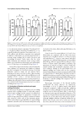
Figure 3. In vitro screening of pMSC donors. (Left to right) Compressive modulus and biochemical contents (DNA, sulfated glycosaminoglycan (sGAG),
and collagen (COL) contents) of composites containing encapsulated adult pMSCs from 3 donors and cultured for 28 days in chondrogenic media. n = 4,
one-way ANOVA with Tukey’s HSD post-hoc test, *p < 0.05, ns = not significant.
n = 6), pinned precultured composites (“Precultured+Pin,” the border of the defect, albeit with some fibrillation at the
n = 4), glued acellular composites (“Acellular+FG,” n = 4), defect interface.
or glued precultured composites (“Precultured+FG,” n = 4). 2
Both acellular and cell-laden composites were implanted Given the size of the created defects (~0.13 cm ), these
in defects to characterize any potential improvements in observations are consistent with the short-term repair
2
resultant repair cartilage due to pMSC preculture and outcomes seen clinically for defects <4 cm that are treated
[41]
neocartilage formation. Twelve weeks after the initial with microfracture . However, some pinned and glued
defect formation and treatment, repair cartilage within composites also exhibited heterogeneous and incomplete
defects was evaluated via arthroscopy, gross observation, defect filling, with more apparent fissures along the defect
micro-CT, histology, and mechanical testing (Figure 5). border in defects treated with pinned composites. The
appearance of repaired cartilage in defects treated with
Across all the surgeries in which precultured composites pinned acellular and precultured composites varied. In
were implanted and explanted, the viability of implants some instances, damage to the subchondral bone and
was first investigated via Live/Dead staining of similarly adjacent cartilage was evident, while in other examples,
fabricated constructs (same cell and material batch, repaired tissue adjacent to the pin was opaque and level with
fabricated at the same time) after the first 7 days of culture the adjacent tissue . While glued composites resulted in
[30]
in chondrogenic media. Importantly, no differences in cell similarly heterogeneous repair cartilage, in more instances,
viability (~79.4% ± 3.0%, n = 21) were observed across glued composites facilitated complete defect filling and the
precultured composites prepared for separate surgeries. In formation of smooth cartilage that integrated with the
addition, the cell viability observed across all these samples surrounding tissue.
was consistent with that previously observed in our donor
screening study (Figure 2C). Macroscopic observation of the femoral trochlea
corroborated the trends observed via arthroscopy.
3.3. Evaluation of fixation methods and repair Interestingly, 8 of the 10 defects treated with pinned
cartilage formation composites contained a visible pinhead after explanting
To characterize the presence of implants and the healing the femoral trochlea; although it is expected that the pin
response within cartilage defects 12 weeks after surgery, would be retained within all defects after 12 weeks based
nascent repair cartilage was first assessed via arthroscopy on their previously reported degradation behavior, a 75%
(Figure 6) [29,40] . Although the appearance of defects and success rate of implant fixation was also previously reported
repair cartilage varied across animals, a number of features with the use of these pins in the same animal model .
[16]
were conserved within each of the treatment groups. Although unlikely, retention of implants within the two
Within defects that were generally filled, the repair cartilage defects where the pin could not be readily seen may still
exhibited a smooth surface and intimate integration along have been achieved. Given that at least one instance of pin
Volume 9 Issue 5 (2023) 500 https://doi.org/10.18063/ijb.775