
Page 66 - IJB-9-5
P. 66
International Journal of Bioprinting 3D-printed PLA-BG composite induces angiogenesis
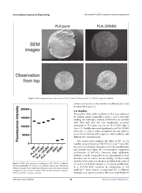
Figure 2. SEM images and gross observation of the 3D-printed PLA pure and PLA–20%BG composite scaffolds.
content, an increase in the number of adhered cells could
be observed (Figure 4).
3.4. Viability
The positive effect of BG inclusion to PLA was confirmed
by viability assays (alamarBlue assay) 1 and 4 days after
seeding. On both days, viability of HUVECs on scaffolds
with 10% and 20% BG was significantly increased
compared to PLA pure and also to PLA-5%BG (day 4;
Figure 5). Viability increased significantly on PLA–20%BG
from day 1 to day 4, which manifested the best effect in
terms of cell viability with respect to other scaffolds with
different BG concentrations.
Few studies have analyzed the effect of BG on the
[26]
viability and proliferation of HUVECs. Li et al. tested BG
ion extracts in different dilutions on HUVECs proliferation
and showed that higher BG concentrations suppressed
proliferation of HUVECs. However, their application
method is hardly comparable to our experiments as their
intention was to induce wound healing. Another study
used BG–PVA (polyvinyl alcohol) scaffolds in the ratios of
Figure 3. MTT tests performed analogous to ISO 10993-5 confirmed 4:1 and 3:1 and demonstrated an increased proliferation
the biocompatibility of all four PLA scaffolds without and with BG in of a HUVEC-hOB coculture in comparison to HA
different concentrations. Significant differences were only observed when [28]
compared to the cytotoxic controls. NM: normal cultivation medium; scaffolds . Some studies incorporated BG in different
ZDEC and ZDBC: cytotoxic controls. hydrogels and reported positive effects on endothelial cell
Volume 9 Issue 5 (2023) 58 https://doi.org/10.18063/ijb.751